
Page 295 - Clinical Manual of Small Animal Endosurgery
P. 295
Small Exotic Animal Endosurgery 283
The cranial kidney, gonad and adrenal gland are visible through the
abdominal air sac, with the ureter and uterus or ductus deferens ventral
to them. Cranial to this the left lung may be visualised, and in larger
birds the endoscope can be advanced into the ostium to examine the
bronchi. The heart, edge of the liver and medial aspect of lung can be
visualised by directing the endoscope cranially into the cranial air-sac
space. The spleen is normally located within the abdominal air sac,
ventral to the kidney, in the junction between proventriculus and gizzard,
but may be obscured in female birds with an active ovary.
Avian endosurgical organ biopsies are extremely small (1–1.5 mm)
when using flexible biopsy forceps via the 2.7 mm endoscope and operat-
ing sheath. While this limits tissue trauma, it can also limit the diagnostic
value unless care is taken. Any visible lesions should be targeted for
sampling, as pathological changes may not be diffusely distributed.
When performing a liver or lung biopsy it is advisable to first incise the
overlying air sac and serosal membranes. This minimises crush artefact
as well as ensuring that hepatic parenchyma is indeed sampled. If pos-
sible more than one biopsy should be taken. Prior use of scissors is not
generally necessary when biopsing the kidney, spleen, adrenal glands and
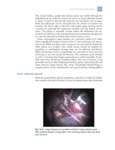
many obvious visual lesions (Fig. 10.6). Post-biopsy haemorrhage is
usually minor in birds, which tend to have good extrinsic clotting ability.
Ventral coelioscopy approach
Birds are positioned in dorsal recumbency, and entry is made on midline
just caudal to the end of the keel. A layer of adipose tissue may be present
Fig. 10.6 Larger biopsies are possible in birds if using multiple ports.
Here a kidney biopsy is being taken. The overlying capsule does not need
prior incision.