
Page 299 - Clinical Manual of Small Animal Endosurgery
P. 299
Small Exotic Animal Endosurgery 287
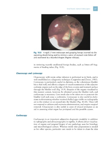
Fig. 10.8 A rigid 2.7 mm endoscope and grasping forceps inserted via the
operating sheath being used to retrieve a piece of stomach tube bitten off
and swallowed by a bearded dragon (Pogona vitticeps).
in retrieving recently swallowed foreign bodies, such as bitten-off frag-
ments of feeding tubes (Fig. 10.8).
Cloacoscopy and cystoscopy
Cloacoscopy with warm saline infusion is performed as in birds, and is
well established as a diagnostic technique (Coppoolse and Zwart, 1985).
Cystoscopy is particularly useful in chelonia, as the voluminous bladder
has a thin wall, and allows a degree of visual examination of the adjacent
coelomic organs such as the edge of the liver, ovaries and stomach surface
through the bladder wall (Fig. 10.9). Biopsies of the organs visualised in
this manner cannot, however, be obtained across the bladder wall, and
coelioscopy is necessary. Care needs also to be taken not to puncture the
bladder wall and cause a coelomitis. Cystoscopy is also useful in chelo-
nians in determining whether retained shelled eggs visible on radiographs
are in the oviduct or are passed into the bladder (Fig. 10.10). These will
not respond to calcium and oxytocin administration, and require surgical
removal. Cloacoscopy is also useful in cases of cloacal prolapses as an
aid in assessing what organ has prolapsed and the possible causes.
Coelioscopy
Coelioscopy is an important adjunctive diagnostic modality in addition
to radiography and ultrasonography in reptiles. It allows direct visualisa-
tion of organs and targeted biopsy of any pathology seen for histology
and microbiological culture. Whereas sterile skin preparation is needed
as for other species, particular care needs to be taken to clean the skin