
Page 305 - Clinical Manual of Small Animal Endosurgery
P. 305
Small Exotic Animal Endosurgery 293
(a) (b)
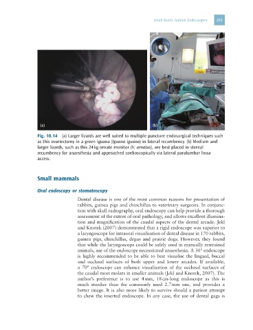
Fig. 10.14 (a) Larger lizards are well suited to multiple puncture endosurgical techniques such
as this ovariectomy in a green iguana (Iguana iguana) in lateral recumbency. (b) Medium and
larger lizards, such as this 24 kg ornate monitor (V. ornatus), are best placed in sternal
recumbency for anaesthesia and approached coelioscopically via lateral paralumbar fossa
access.
Small mammals
Oral endoscopy or stomatoscopy
Dental disease is one of the most common reasons for presentation of
rabbits, guinea pigs and chinchillas to veterinary surgeons. In conjunc-
tion with skull radiography, oral endoscopy can help provide a thorough
assessment of the extent of oral pathology, and allows excellent illumina-
tion and magnification of the caudal aspects of the dental arcade. Jekl
and Knotek (2007) demonstrated that a rigid endoscope was superior to
a laryngoscope for intraoral visualisation of dental disease in 170 rabbits,
guinea pigs, chinchillas, degus and prairie dogs. However, they found
that while the laryngoscope could be safely used in manually restrained
animals, use of the endoscope necessitated anaesthesia. A 30° endoscope
is highly recommended to be able to best visualise the lingual, buccal
and occlusal surfaces of both upper and lower arcades. If available,
a 70° endoscope can enhance visualisation of the occlusal surfaces of
the caudal most molars in smaller animals (Jekl and Knotek, 2007). The
author’s preference is to use 4 mm, 18 cm-long endoscope as this is
much sturdier than the commonly used 2.7 mm one, and provides a
better image. It is also more likely to survive should a patient attempt
to chew the inserted endoscope. In any case, the use of dental gags is