Page 310 - Clinical Manual of Small Animal Endosurgery
P. 310
298   Clinical Manual of Small Animal Endosurgery


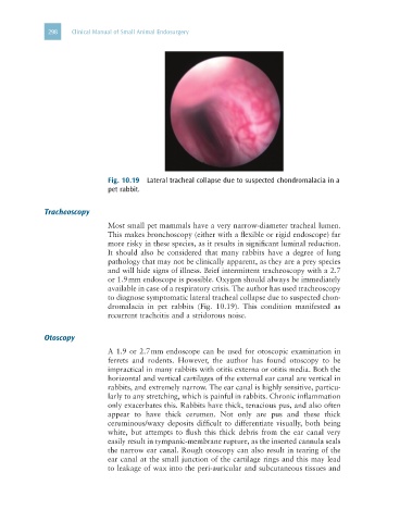
                              Fig. 10.19  Lateral tracheal collapse due to suspected chondromalacia in a
                              pet rabbit.


             Tracheoscopy
                              Most small pet mammals have a very narrow-diameter tracheal lumen.
                              This makes bronchoscopy (either with a flexible or rigid endoscope) far
                              more risky in these species, as it results in significant luminal reduction.
                              It should also be considered that many rabbits have a degree of lung
                              pathology that may not be clinically apparent, as they are a prey species
                              and will hide signs of illness. Brief intermittent tracheoscopy with a 2.7
                              or 1.9 mm endoscope is possible. Oxygen should always be immediately
                              available in case of a respiratory crisis. The author has used tracheoscopy
                              to diagnose symptomatic lateral tracheal collapse due to suspected chon-
                              dromalacia  in  pet  rabbits  (Fig.  10.19).  This  condition  manifested  as
                              recurrent tracheitis and a stridorous noise.


             Otoscopy
                              A 1.9 or 2.7 mm endoscope can be used for otoscopic examination in
                              ferrets  and  rodents.  However,  the  author  has  found  otoscopy  to  be
                              impractical in many rabbits with otitis externa or otitis media. Both the
                              horizontal and vertical cartilages of the external ear canal are vertical in
                              rabbits, and extremely narrow. The ear canal is highly sensitive, particu-
                              larly to any stretching, which is painful in rabbits. Chronic inflammation
                              only exacerbates this. Rabbits have thick, tenacious pus, and also often
                              appear  to  have  thick  cerumen.  Not  only  are  pus  and  these  thick
                              ceruminous/waxy deposits difficult to differentiate visually, both being
                              white,  but  attempts  to  flush  this  thick  debris  from  the  ear  canal  very
                              easily result in tympanic-membrane rupture, as the inserted cannula seals
                              the narrow ear canal. Rough otoscopy can also result in tearing of the
                              ear canal at the small junction of the cartilage rings and this may lead
                              to leakage of wax into the peri-auricular and subcutaneous tissues and

