
Page 379 - Veterinary Histology of Domestic Mammals and Birds, 5th Edition
P. 379
Receptors and sense organs (organa sensuum) 361
mentation of the retinal pigment epithelium is markedly 16.20 to 16.23). As a component of the diencephalon, this
VetBooks.ir reduced or absent. part of the retina contains glial cells (Müller cells, radial
Additional functions performed by the retinal pigment glial cells) and nerve cells (neurons).
Müller cells (Figures 16.23 and 16.26) support all layers
epithelium include:
of the neurosensory retina except the rods and cones. They
· nutritional support for the retina, also form the internal and external limiting membranes
· transport of metabolites between the choroid and (stratum limitans gliae internum and externum).
the outer layers of the sensory retina, The neurons are arranged in structurally distinct layers
· uptake and digestion of phagocytosed material that are connected in series to form a three-neuron path-
from the outer segments (membranous discs) of the way. From outside (most external) to inside (most internal)
photoreceptors, (Figures 16. 20 to 16.23) the pathway consists of:
· replenishment of rhodopsin through esterification
of vitamin A and · neuron 1:
· synthesis of melanin. − rod and cone layer (stratum neuro-epitheliale),
− outer nuclear layer (stratum nucleare externum),
NEUROSENSORY RETINA (STRATUM NERVOSUM − outer plexiform layer (stratum plexiforme
RETINAE) externum),
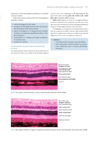
The neurosensory retina, derived from the inner leaf of
the embryonic optic cup, consists of several layers (Figures
16.20 Pars optica retinae (fundus, horse). Haematoxylin and eosin stain (x250).
16.21 Pars optica retinae in region of tapetum lucidum (fundus, horse). Haematoxylin and eosin stain (x250).
Vet Histology.indb 361 16/07/2019 15:07