Page 170 - Manual of Equine Field Surgery
        P. 170
     166                        OPHTHALMIC  SURGERIES
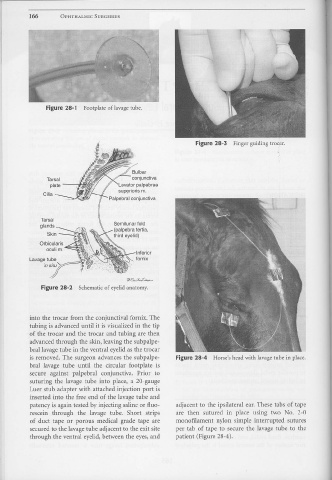
                                        Figure  28-1                   Footplate  of lavage tube.
                                                                                                                                                                                                                                             •
                                                                                                                                                                      Figure 28-3  Finger  guiding  trocar .
                                                                                                                                                                                                                                            •  .
                                                                                                                                                                                                                                           '
                                        Tarsal                                                                  conjunctiva
                                           plate                                                     Levator  palpebrae
                                                                                                     superloris  m.
                                     Cilia
                                                                                             Palpebral  conjunctiva
                                   Tarsal
                                                                                                 Semilunar  fold
                                   glands  ----                                                  (palpebra  tertia,
                                         Skin             ---.0
                                                                                                 third  eyelid)
                                                                                                       :.u.\--      Inferior
                          Lavage  tube                  :;:;::=~                                                    fornix
                                      in  situ"'-...-::
                                                                                                            ~~;t-,..,....
                                    Figure  28-2  Schematic  of eyelid anatomy.
                          into  the trocar  fron1 the  conjunctiva! fornix. The
                          tubing  is advanced until  it is visualized i11 the tip
                          of the  trocar  and  the  trocar  and  tubing  are then
                          advanced through  the skin, leaving the subpalpe-
                          bral lavage tube in the ventral  eyelid as the trocar
                          is removed.  The  surgeon  advances the  subpalpe-                                                                         Figure 28-4  Horse's head with lavage tube  in place.
                          bral  lavage  tube  until  the  circular  footplate  is
                          secure  against  palpebral  conjunctiva.  Prior  to
                          suturing  the  lavage tube  into  place,  a  20-gauge
                          Luer stub  adapter  with  attached  injection  port is
                          inserted  into  the  free end  of the lavage tube  and
                          patency is again tested by injecting saline or fluo-                                                                       adjacent to  the  ipsilateral  ear. These tabs  of tape
                          rescein  through  the  lavage  tube.  Short  strips                                                                        are  then  sutured  in  place  using  two  No.  2-0
                          of duct  tape  or  porous  medical  grade  tape  are                                                                       monofilament  nylon  simple  interrupted  sutures
                          secured to the lavage tube adjacent to the exit site                                                                       per  tab  of tape  to  secure  the  lavage tube  to  the
                          through  the ventral  eyelid, between the  eyes, and                                                                       patient  (Figure 28-4).