Page 173 - Manual of Equine Field Surgery
        P. 173
     Eyelid  Laceration Repair                                      169
                                                                                                                                             COMPLICATIONS
                                                                                                                                             The  most  frequent  complication  of eyelid lacera-
                                                                                                                                             tion  repair  is  wound  dehiscence.  This  is  most
                                                                                                                                             commonly  a result  of single-layer  closure,  but  it
                                                                                                                                             may  also  occur  as a  result  of  devitalized  wound
                                                                                                                                             edges or  excessive tension  across the  surgical  site.
         A                                                                   B                                                               If  wound  dehiscence  occurs,  a  second  surgical
                                                                                                                                              repair  with  debridement  and  suturing  is  recom-
                                                                                                                                              mended  to  prevent  eyelid  margin  defects  and
                                                                                                                                              to  reduce  the  potential  for  corneal  abrasion  or
                                                                                                                                              ulceration.
                                                                                                                                                    Some patients  may develop  a notch-like  defect
                                                                                                                                              of the eyelid. Such defects, if significant,  can cause
                                                                                                                                              abnormal  tearfilm  distribution,  and  corneal  irri-
                                                                                                                                              tation  or  ulceration.  Minor  defects  of the  eyelid
                                                                                                                                              margin  may be left  alone  if they  do not adversely
                                                                                                                                              affect the corneal surface;  more  significant  defects,
          C                                                                   D                                                               however, require  additional  blepharoplastic  tech-
                                                                                                          ~t?.r,v,f.<. ..:t~--                niques  to  correct  or  remove  the  defect.  These
                                                                                                                                              eyelid margin  defects are most  easily prevented by
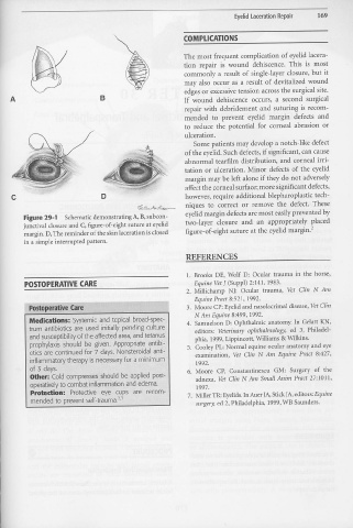
                   Figure 29-1                    Schematic  demonstrating  A, B, subcon-
                   junctival  closure  and C, figure-of-eight  suture at eyelid                                                               two-layer  closure  and  a.11  appropriately  placed
                   margin.  D, The  reminder  of the skin laceration  is closed                                                               figure-of-eight  suture  at the eyelid margin.2
                   in a simple  interrupted  pattern.
                                                                                                                                               REFERENCES
                                                                                                                                               1.  Brooks  DE,  Wolf  D:  Ocular  trauma  in  the  horse,
                                                    I
                    POSTOPERATVE  CARE                                                                                                               Equine  Vet J (Suppl)  2:141,  1983.
                                                                                                                                               2.  Millichamp  NJ:  Ocular  trauma,  Vet  Clin  N  Am
                                                                                                                                                     Equine  Pract 8:521,  1992.
                                 . .  .
                       Postoperative Care "'                                ··.· ..                                                            3.  Moore  CP: Eyelid  and  nasolacrimal  disease,  Vet Clin
                                                                                                                                                     N Am  Equine  8:499,  1992.
                       Medications: Systemic  and  topical  broad-spec-                                                                        4.  Samuelson  D:  Ophthalmic  anatomy.  In  Gelatt  KN,
                       trum  antibiotics  are  used  initially  pending  culture                                                                      editors:  Veterinary  ophthalmology,  ed  3,  Philadel-
                       and susceptibility  of the  affected area, and tetanus.                                                                        phia,  1999, Lippincott,  Williams  & Wilkins.
                        prophylaxis  should  be  given.  Appropriate  antibi-                                                                   5.  Cooley  PL: Normal  equine  ocular  anatomy  and  eye
                        otics  are  continued  for  7 days.  Nonsteroidal  anti-                                                                      examination,  Vet  Cli11  N  Am  Equine  Pract  8:427,
                        inflammatory  therapy  is necessary for  a  minimum                                                                           1992.
                        of  3 clays.                                                                                                            6.  Moore  CP,  Constantinescu                                    GM:  Surgery  of  the
                        other: Cold  compresses  should  be  applied  post-                                                                           adnexa,  Vet Clin  N Am  Small  Anim  Pract  27:1011,
                        operatively to combat  inflammation  and  edema.                                                                              1997.
                        Protedion:  Protective  eye  cups  are  recom-                                                                          7.  Miller TR:  Eyelids. In Auer JA, Stick JA, editors: Equin.e
                        mended  to  prevent  self-trauma.2•3                                                                                          surgery, ed 2, Philadelphia,  1999, WB Saunders.


