Page 176 - Manual of Equine Field Surgery
P. 176
172 OPHTHALMIC SURGERIES
POSTOPERATIVE CARE
r . 1 • c
r,,ostop.eratve are
Protedion: A protective evecup is used for 1
A week postoperatively to prevent self-trauma.
Medications: Nonsteroidal antiinflammatory
drugs may be used from 3 to 7 days to minimize
associated discomfort and edema.3
B Suture removal: Suture removal is recom- •
mended in approximateJy 14 days.
COMPLICATIONS
c
I
' --- .,.."" ,
\ I Patients commonly exhibit significant periorbital
<,
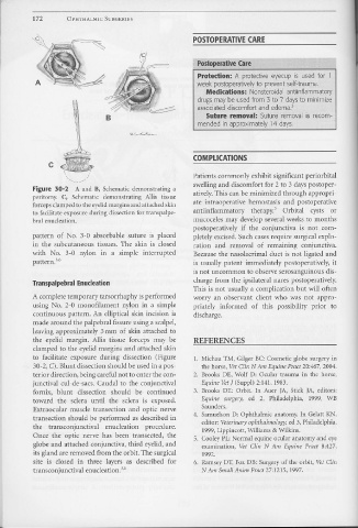
Figure 30-2 A and B, Schematic demonstrating a swelling and discomfort for 2 to 3 days postoper-
peritomy. C, Schematic demonstrating Allis tissue atively. This can be minimized through appropri-
forceps clamped to the eyelid margins and attached skin ate intraoperative hemostasis and postoperative
to facilitate exposure during dissection for transpalpe- antiinflammatory therapy. 3 Orbital cysts or
bral enucleation. mucoceles may develop several weeks to months
postoperatively if the conjunctiva is not com-
pattern of No. 3-0 absorbable suture is placed pletely excised. Such cases require surgical explo-
in the subcutaneous tissues. The skin is closed ration and removal of remaining conjunctiva.
with No. 3-0 nylon in a simple interrupted Because the nasolacrimal duct is not ligated and
pattern. 3'6 is usually patent immediately postoperatively, it
is not uncommon to observe serosanguinous dis-
Transpalpebral Enucleation charge from the ipsilateral nares postoperatively.
This is not usually a complication but will often
A complete temporary tarsorrhaphy is performed worry an observant client who was not appro-
using No. 2-0 monofilament nylon in a simple priately informed of this possibility prior to
continuous pattern. An elliptical skin incision is discharge.
made around the palpebral fissure using a scalpel,
leaving approximately 5 mm of skin attached to
the eyelid margin. Allis tissue forceps may be REFERENCES
clamped to the eyelid margins and attached skin
to facilitate exposure during dissection (Pigure 1. Michau TM, Gilger BC: Cosmetic globe surgery in
30-2, C). Blunt dissection should be used in a pos- the horse, Vet Clin N Am Equine Pract 20:467, 2004.
terior direction, being careful not to enter the con- 2. Brooks DE, Wolf D: Ocular trauma in the horse,
junctiva! cul-de-sacs. Caudal to the conjunctiva! Equine Vet J (Suppl) 2:141, 1983.
fornix, blunt dissection should be continued 3. Brooks DE: Orbit. In Auer JA, Stick JA, editors:
toward the sclera until the sclera is exposed. Equine surge1y, ed 2, Philadelphia, 1999, WB
Extraocular muscle transection and optic nerve Saunders.
transection should be performed as described in 4. Samuelson D: Ophthalmic anatomy. In Gelatt KN,
editor: Veterinary ophthalmology, ed 3, Philadelphia,
the transconjunctival enucleation procedure. 1999, Lippincott, Williams & Wilkins.
Once the optic nerve has been transected, tl1e 5. Cooley PL: Normal equine ocular anatomy and eye
globe and attached conjunctiva, third eyelid, and examination, Vet Clin N Am Equine Pract 8:427,
its gland are removed from the orbit. The surgical 1992.
site is closed in three layers as described for 6. Ramsey DT, Fox DB: Surgery of the orbit, Vet Clin
transconjunctival enucleation. 3'6 N Am Small Anim Pract 27:1215, 1997.

