Page 210 - Manual of Equine Field Surgery
P. 210
CHAPTER                                                                   37


                                                                                                            Penile Amputation


                                                                                                                       Joanne  Kramer


                                                                                                                                                     nosum  penis  surrounded  by the tunica albuginea
                          INDICATIONS                                                                                                                is the  largest  vascular  space.  The  primary blood


                                                                                                                                                     vessels ( the  dorsal  arteries  and  veins  of the  penis)

                          Neoplastic  lesions  (primarily squamous  cell carci-                                                                       encountered  in  penile  amputation  are  on  the


                          noma)  and  other  masses involving the shaft of the                                                                        dorsal  aspect  of the penis between the deep fascia


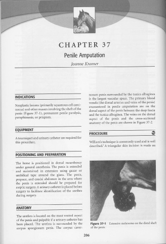
                          penis  (Figure  3 7-1),  permanent  penile  paralysis,                                                                      and  the tunica  albuginea.  The veins on the  dorsal


                          paraphimosis, or priapism.                                                                                                  aspect  of  the  penis  and  the  cross-sectional


                                                                                                                                                      anatomy  of the penis  are shown  in Figure  37-2.


                           EQUIPMENT


                                                                                                                                                      PROCEDURE


                           A tourniquet  and urinary catheter  are required  for


                           this procedure.                                                                                                            William's technique  is commonly  used and is well

                                                                                                                                                                             1
                                                                                                                                                      described.  A triangular  skin  incision  is made  on


                           POSITIONING AND  PREPARATION


                           The  horse  is  positioned  in  dorsal  recumbency


                           under  general  anesthesia.  The  penis  is  extended


                           and  maintained  in  extension  using  gauze  or


                           umbilical  tape  around  the  glans.  The  penis,


                           prept1ce, and  caudal  abdomen  in  the  area  where


                           the  penis  is  extended  should  be  prepared  for


                            aseptic surgery. A urinary  catheter  is placed before


                            surgery  to  facilitate  identification  of  the  urethra


                            during  surgery.


                            ANATOMY


                            The urethra is located  on  the  most  ventral  aspect


                            of the penis  and palpable if a urinary catheter  has


                            been  placed.  The  urethra  is  surrounded  by  the                                                                        Figure  37-1                   Extensive melanoma  on the distal shaft


                             corpus  spongiosum  penis.  The  corpus  caver-                                                                            of the penis.


                                                                                                                                            206

