Page 212 - Manual of Equine Field Surgery
P. 212
208                       MALE  UROGENITAL SURGERIES

  I                                                                                                                                                                                                                                                                                  •


                                                                                                                                                          .
                                                                                                                                                          .                                                                                                                           I


                                                                                                                                                                                                                                                                                      f


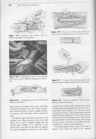
                                                                                                                                                      Figure  37-6  The  sides  of  the  incised  urethra  are


                                                                                                                                                      sutured  to the  epithelium  using  an interrupted pattern.

                           Figure  37-3  Triangular                                    skin  incision  used  in


                           William's  technique  for amputation.                                                                                                                                                              .,,.,_   .flJC.~


                                                                                                                                                                                                                              .·,-·


                            Figure  37-4  Intraoperatjve  view  of  the  triangular


                            skin  incision  used  i11  William's  technique  for  amputa-

                              .
                            tion.                                                                                                                       Figure 37-7  The base of the penis  is transected  so that
                                                                                                                                                        the dorsal aspect is slightly longer than the ventral  aspect.


                             Figure  37-5  A longitudinal  incision  is made  into  the                                                                  Figure  37-8  The  tunica  albuginea  is closed  to com-


                             lumen of the urethra.                                                                                                      press  the  corpus  cavernosurn.


                             thral  mucosa,  minimizes  dead  space,  and  helps                                                                         cavernosum  to compress  the vascular  spaces using


                              control  hemorrhage  from  the  corpus  spongio-                                                                           a simple  interrupted  pattern  with  No.  0 or No.  1


                              sum. When  this  layer is closed,  a simple  continu-                                                                      absorbable  suture  (Figure  37-8).  The  first  suture


                              ous  pattern  with  absorbable  suture  material  is                                                                       is placed  on  rnidline,  and  the  subsequent  sutures

                                                                                                                                                         bisect the halves (Figure  3 7-9).  The sutures  should
                              used.


                                    The  penis  is then  transected  at the base  of the                                                                 be closely spaced.  Generally, seven or eight sutures


                              triangle  i11 an  oblique  manner  so that  the  dorsal                                                                    are used.  Blood loss from  the  corpus  cavernosum


                              aspect  of  the  penis  is  slightly  longer  than  the                                                                    can be  extensive, and  this  seal should  be  checked


                              ventral  aspect  (Figure  3 7 - 7).  Branches  of  the                                                                     carefully after release  of the tourniquet.


                              dorsal  artery  and  veins  of  the  penis  are ligated.                                                                          The  subcutaneous  tissue  deep  to  the  skin  is


                              The  tunica  albuginea  is  closed  over  the  corpus                                                                       closed  to  the  tissue  just  deep  to  the  urethral

