
Page 13 - Basic Monitoring in Canine and Feline Emergency Patients
P. 13
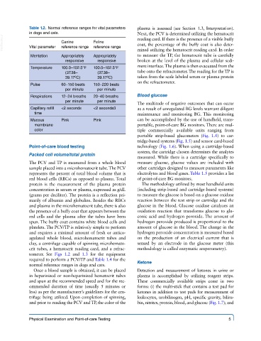
Table 1.2.  Normal reference ranges for vital parameters   plasma is assessed (see Section 1.3, Interpretation).
             in dogs and cats.                           Next, the PCV is determined utilizing the hematocrit
  VetBooks.ir  Vital parameter  Canine    Feline         reading card. If there is the presence of a visible buffy
                                                         coat, the percentage of the buffy coat is also deter-
                          reference range
                                       reference range
                                                         mined utilizing the hematocrit reading card. In order
              Mentation   Appropriately   Appropriately   to measure the TP, the hematocrit tube is carefully
                            responsive   responsive      broken at the level of the plasma and cellular sedi-
                                                         ment interface. The plasma is then evacuated from the
              Temperature  100.0–102.5°F   100.0–102.5°F
                            (37.38–      (37.38–         tube onto the refractometer. The reading for the TP is
                            39.17ºC)     39.17ºC)        taken from the scale labeled serum or plasma protein
                                                         on the refractometer.
              Pulse       60–150 beats    150–220 beats
                            per minute   per minute
              Respirations  12–24 breaths    20–40 breaths   Blood glucose
                            per minute   per minute      The multitude of negative outcomes that can occur
              Capillary refill    <2 seconds  <2 seconds3  as a result of unregulated BG levels warrant diligent
               time                                      maintenance and  monitoring  BG.  This  monitoring
              Mucous      Pink         Pink              can be accomplished by the use of handheld, trans-
               membrane                                  portable, point-of-care BG monitors. There are mul-
               color                                     tiple commercially available units ranging from
                                                         portable  strip-based  glucometers  (Fig.  1.4)  to  car-
                                                         tridge-based systems (Fig. 1.5) and sensor card-based
             Point-of-care blood testing                 technology (Fig. 1.6). When using a cartridge-based
                                                         system, the cartridge chosen determines the analytes
             Packed cell volume/total protein
                                                         measured. While there is a cartridge specifically to
             The PCV and TP is measured from a whole blood   measure glucose, glucose values are included with
             sample placed into a microhematocrit tube. The PCV   other cartridges designed to measure parameters like
             represents the percent of total blood volume that is   electrolytes and blood gases. Table 1.5 provides a list
             red blood cells (RBCs) as opposed to plasma. Total   of point-of-care BG monitors.
             protein is the measurement of the plasma protein   The methodology utilized by most handheld units
             concentration in serum or plasma, expressed as g/dL   (including strip-based and cartridge-based systems)
             (grams per deciliter). The protein is a reflection pri-  to measure the glucose is based on a glucose oxidase
             marily of albumin and globulins. Besides the RBCs   reaction between the test strip or cartridge and the
             and plasma in the microhematocrit tube, there is also   glucose in the blood. Glucose oxidase catalyzes an
             the presence of a buffy coat that appears between the   oxidation reaction that transforms glucose to glu-
             red cells and the plasma after the tubes have been   conic acid and hydrogen peroxide. The amount of
             spun. The buffy coat contains white blood cells and   hydrogen peroxide produced is proportional to the
             platelets. The PCV/TP is relatively simple to perform   amount of glucose in the blood. The change in the
             and requires a minimal amount of fresh or antico-  hydrogen peroxide concentration is measured based
             agulated whole blood, microhematocrit tubes and   on the production of an electrical current that is
             clay, a centrifuge capable of spinning microhemato-  sensed by an electrode in the glucose meter (this
             crit tubes, a hematocrit reading card, and a refrac-  methodology is called enzymatic amperometry).
             tometer. See  Figs 1.2 and  1.3 for the equipment
             required to perform a PCV/TP and Table 1.4 for the
             normal reference ranges in dogs and cats.   Ketone
               Once a blood sample is obtained, it can be placed   Detection and measurement of ketones in urine or
             in heparinized  or non-heparinized hematocrit tubes   plasma is accomplished by utilizing reagent strips.
             and spun at the recommended speed and for the rec-  These commercially available strips come in two
             ommended duration of time (usually 5 minutes or   forms: (i) the multi-stick that contains a test pad for
             less) as per the manufacturer’s guidelines for the cen-  ketones in addition to test pads for measurement of
             trifuge being utilized. Upon completion of spinning,   leukocytes, urobilinogen, pH, specific gravity, biliru-
             and prior to reading the PCV and TP, the color of the   bin, nitrites, protein, blood, and glucose (Fig. 1.7); and


             Physical Examination and Point-of-care Testing                                    5