
Page 168 - Basic Monitoring in Canine and Feline Emergency Patients
P. 168
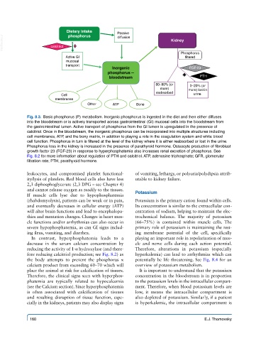
Dietary intake       Passive
                             phosphorus
  VetBooks.ir         calcitriol                                       Kidney
                                                 diffusion
                                                                           Phosphorus
                            Active GI                                        filtered
                            mucosal
                            transport
                                               Inorganic                       FGF-23
                                             phosphorus --
                                              bloodstream                          PTH
                                                                 80–90% (or    0–20% (or
                                                                   more)      more) lost in
                                                                 reabsorbed
                          Cell                                                  urine
                        membranes
                                     Other     ATP       Bone

            Fig. 8.3.  Basic phosphorus (P) metabolism. Inorganic phosphorus is ingested in the diet and then either diffuses
            into the bloodstream or is actively transported across gastrointestinal (GI) mucosal cells into the bloodstream from
            the gastrointestinal lumen. Active transport of phosphorus from the GI lumen is upregulated in the presence of
            calcitriol. Once in the bloodstream, the inorganic phosphorus can be incorporated into multiple structures including
            cell membranes, ATP, and the bony matrix, in addition to playing a role in the coagulation system and white blood
            cell function. Phosphorus in turn is filtered at the level of the kidney where it is either reabsorbed or lost in the urine.
            Phosphorus loss in the kidney is increased in the presence of parathyroid hormone. Osteocyte production of fibroblast
            growth factor 23 (FGF-23) in response to hyperphosphatemia also increases renal excretion of phosphorus. See
            Fig. 8.2 for more information about regulation of PTH and calcitriol. ATP, adenosine triphosphate; GFR, glomerular
            filtration rate; PTH, parathyroid hormone.


            leukocytes, and compromised platelet functional-  of vomiting, lethargy, or polyuria/polydipsia attrib-
            ity/lysis of platelets. Red blood cells also have less   utable to kidney failure.
            2,3 diphosphoglycerate (2,3 DPG – see Chapter 4)
            and cannot release oxygen as readily to the tissues.
            If  muscle cells lyse due to hypophosphatemia   Potassium
            (rhabdomyolysis), patients can be weak or in pain,   Potassium is the primary cation found within cells.
            and eventually decreases in cellular energy (ATP)   Its concentration is similar to the extracellular con-
            will alter brain functions and lead to encephalopa-  centration of sodium, helping to maintain the elec-
            thies and mentation changes. Changes in heart mus-  trochemical balance.  The majority of potassium
            cle functions and/or arrhythmias can also occur in   (66–75%) is contained within muscle cells.  The
            severe hypophosphatemia, as can GI signs includ-  primary role of potassium is maintaining the rest-
            ing ileus, vomiting, and diarrhea.           ing membrane potential of the cell, specifically
              In contrast, hyperphosphatemia leads to a   playing an important role in repolarization of mus-
            decrease in the serum calcium concentration by   cle and nerve cells during each action potential.
            reducing the activity of 1-α hydroxylase (and there-  Therefore, alterations in potassium (especially
            fore reducing calcitriol production; see Fig. 8.2) as   hyperkalemia) can lead to arrhythmias which can
            the body attempts to prevent the phosphorus ×   potentially be life threatening. See Fig. 8.4 for an
            calcium product from exceeding 60–70 which will   overview of potassium metabolism.
            place the animal at risk for calcification of tissues.   It is important to understand that the potassium
            Therefore, the clinical signs seen with hyperphos-  concentration in the bloodstream is in proportion
            phatemia  are typically related to  hypocalcemia   to the potassium levels in the intracellular compart-
            (see the Calcium section). Since hyperphosphatemia   ment. Therefore, when blood potassium levels are
            is  often  associated  with  calcification  of  tissues   low,  it  means  the  intracellular  compartment  is
            and  resulting disruption of tissue function, espe-  also depleted of potassium. Similarly, if a patient
            cially in the kidneys, patients may also display signs   is  hyperkalemic, the intracellular compartment is


             160                                                                     E.J. Thomovsky