
Page 169 - Basic Monitoring in Canine and Feline Emergency Patients
P. 169
Dietary intake
VetBooks.ir
Potassium in Catecholamines Potassium into
Passive absorption bloodstream/ Translocation cells
stomach and small interstitium Insulin
intestine
Excreted Hyperkalemia
through Hypokalemia Acidemia Alkalemia
+
+
colon (–) (↑ H ) (↓ H )
↑ Aldosterone H into cells H into blood
+
+
↑ colonic secretion Excreted through K into blood K into cells
+
+
(loss) of K + kidney
+
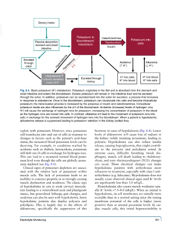
Fig. 8.4. Basic potassium (K ) metabolism. Potassium originates in the diet and is absorbed from the stomach and
small intestine and enters the bloodstream. Excess potassium will remain in the intestinal tract and be excreted
through the colon. In addition, potassium can be secreted back into the colon for excretion, a process that increases
in response to aldosterone. Once in the bloodstream, potassium can translocate into cells and become intracellular
potassium; the translocation process is increased by the presence of insulin and catecholamines. Intracellular
potassium levels are also influenced by the pH of the bloodstream. Acidemia (increased levels of hydrogen ions,
+
H ) will cause the exchange of hydrogen ions for potassium, increasing the concentration of potassium in the blood
as the hydrogen ions are moved into cells. In contrast, alkalemia will lead to the movement of potassium ions into
cells in exchange for the outward movement of hydrogen ions into the bloodstream. When a patient is hypokalemic,
aldosterone release is suppressed leading to potassium retention in the kidney (dotted line).
replete with potassium. However, since potassium hormone in cases of hypokalemia (Fig. 8.4). Lower
will translocate into and out of cells in response to levels of aldosterone will cause loss of sodium in
changes in factors such as the patient’s acid–base the kidney (while retaining potassium), leading to
status, the measured blood potassium levels can be polyuria. Hypokalemia can also reduce insulin
deceiving. For example, in conditions marked by release, causing hyperglycemia, that might contrib-
acidemia such as diabetic ketoacidosis, potassium ute to the polyuria and polydipsia noted. In
will shift out of cells in exchange for hydrogen ions. extreme cases, difficulty breathing (weak dia-
This can lead to a measured normal blood potas- phragm), muscle cell death leading to rhabdomy-
sium level even though the cells are globally potas- olysis, and even electrocardiogram (ECG) changes
sium depleted (see Fig. 8.4). can occur. These electrical changes can make
Clinical signs of potassium depletion are associ- hypokalemic patients with arrhythmias more
ated with the relative lack of potassium within refractory to treatment, especially with class I anti-
muscle cells. The lack of potassium leads to an arrhythmics (e.g. lidocaine). Hypokalemia does not
inability to contract properly or as strongly causing usually cause observed clinical signs until K levels
+
muscle dysfunction and weakness. The classic sign are significantly less than 3.0 mEq/L.
of hypokalemia in cats is weak cervical muscula- Hyperkalemia also causes muscle weakness (usu-
ture leading to a ventroflexed neck and plantigrade ally K levels >7.5–8.0 mEq/L). When an animal is
+
stance, but generalized lethargy, gait changes, and hyperkalemic, its cell membranes are initially more
weakness can also be seen with hypokalemia. Some excitable than in a normal setting since the resting
hypokalemic patients also display polyuria and membrane potential of the cells is higher (more
polydipsia. This is largely due to the effects of positive) than at normal potassium levels. In car-
aldosterone, specifically the suppression of this diac muscle cells, this initial hyperexcitability is
Electrolyte Monitoring 161