
Page 1068 - Clinical Small Animal Internal Medicine
P. 1068
1006 Section 9 Infectious Disease
Therapy
VetBooks.ir Clindamycin at 10–12 mg/kg PO q8h for at least one
month is the main drug used for the treatment of canine
neosporosis. Trimethoprim‐sulfa at 15 mg/kg PO q12h
alone or in combination with pyrimethamine at 1 mg/kg
PO q24h for at least a month is another treatment option.
Pyrimethamine may be combined also with clindamycin
at the same doses. Ponazuril has been shown to be effec-
tive in cattle and mice but there are insufficient data on
its efficacy against canine neosporosis. Treatment of
neosporosis may only be partially effective or not
effective at all and long‐term treatment of two months
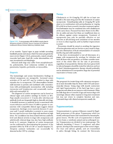
Figure 110.2 A young puppy with hindlimb paresis due to or more is often required, until no further improvement
Neospora caninum infection. Source: Courtesy of Dr Monica is noted.
Leszkowicz Mazuz, Kimron Veterinary Institute, Israel.
Prevention should be aimed at avoiding the ingestion
of bovine placentas and raw bovine or prey meat by dogs,
and also elimination of fecal contamination of ruminant
of six months. Typical signs in pups include ascending
hindlimb paresis and ataxia which becomes progressively feed by dog and wild canid feces.
more severe (Figure 110.2), polymyositis, muscle atrophy, It has been recommended to test all littermates of a
muscular back pain, head tilt, ocular abnormalities, uri- puppy with neosporosis by serology for infection and
nary incontinence and dysphagia. treat all those who are positive, or further consider treat-
Infected adult dogs may suffer from encephalomyeli- ment of the whole litter for the sake of prevention,
tis, polymyositis, focal cutaneous nodules or ulcers, regardless of seropositivity. Bitches who have given birth
pneumonia, hepatitis, peritonitis, or myocarditis. to infected puppies should be tested for infection and not
be bred. Immunosuppressive therapy should preferably
not be administered to seropositive dogs due to potential
Diagnosis reactivation of tissue cysts.
The hematologic and serum biochemistry findings in
clinical neosporosis are nonspecific. Increased serum Prognosis
activities of CK and AST may be evident in dogs with
myositis due to neosporosis. CSF abnormalities include The prognosis for treated dogs with cutaneous neosporo-
mild increase in protein and nucleated cells concentra- sis is more favorable than for those with CNS and neuro-
tions with predominantly mononuclear cells including muscular disease. Young dogs with muscle contracture
monocytes and lymphocytes and occasionally neutro- and rigid hyperextension of the back legs have a poor
phils and eosinophils [6]. prognosis and often do not improve with treatment. Older
The diagnosis of canine neosporosis can be based on juvenile dogs above 4 months of age and mature dogs with
clinical signs and positive serology by ELISA or IFA. neurologic disease often respond to long‐term treatment
Pups usually seroconvert 2–3 weeks after infection and but may not return to complete function and cure.
antibody levels are frequently high in clinically sick dogs. Untreated dogs with clinical neosporosis usually die.
A fourfold increase in antibody levels is associated with
recent infection and IFA titers of 1:800 or greater in con-
junction with compatible clinical signs suggest clinical Trypanosomiasis
neosporosis. Serologic cross‐reactivity between N. cani-
num and T. gondii antibodies is present in some assays Trypanosomiasis is a group of diseases caused by flagel-
but not expected in sera from dogs with medium to high lated protozoans of the genus Trypanosoma which are
titers. No correlation has been found between antibody mostly arthropod borne and transmitted by hematopha-
level and clinical severity in dogs with neosporosis and geous insects. The life cycle of trypanosomes is shared
subclinically infected dogs may also have high antibody by two hosts: a vertebrate animal and an arthropod vec-
levels to N. caninum. Suspicion of canine neosporosis tor. Trypanosomes are divided to two main groups. The
can be confirmed by demonstrating the presence of the Salivaria (including T. brucei, T. conglonese, and other
parasite’s DNA by PCR of CSF or muscle biopsies [7]. species, develop in the anterior part of the vector’s diges-
Neospora caninum tachyzoites can occasionally be tive tract and salivary glands, and are transmitted
detected in the CSF by cytology. in saliva through biting. In contrast the Stercocaria