
Page 1066 - Clinical Small Animal Internal Medicine
P. 1066
1004 Section 9 Infectious Disease
History and Clinical Signs with centrifugation using sugar solutions such as Sheather’s
VetBooks.ir Clinical signs in cats are rarely related to the gastrointes- sugar solution. Hammondia hammondi and Besnoitia
spp. produce oocysts which are morphologically indis-
tinal stage of T. gondii development which is usually sub-
clinical. Cats affected by systemic infection may show a tinguishable from T. gondii occysts in cat feces.
Diagnosis of clinically ill cats and dogs is based on
variety of clinical signs including fever, anorexia, abdom- compatible clinical signs and laboratory abnormalities
inal pain, respiratory distress, ocular inflammation, leth- and by the detection of specific serum antibodies, dem-
argy, and central nervous system disorders. These are onstration of parasite DNA by PCR, or visualization of
associated with tachyzoite dissemination and their cyto- tachyzoites by cytology in clinical samples. T. gondii tach-
toxicity with generation of local and systemic inflamma- yzoites may be detected by cytology during acute toxo-
tion. Acute disease in dogs may be accompanied by plasmosis in peritoneal and ascitic fluids, and more rarely
neuromuscular, central nervous system (CNS), respira- in cerebrospinal fluid (CSF), blood and bronchoalveolar
tory, gastrointestinal, hepatic, and more rarely ocular lavage washings or other tissues. PCR performed on CSF,
manifestations. Myositis and myocarditis are also preva- aqueous humor, or any other body fluid or internal organ
lent in canine toxoplasmosis. Disease in both cats and tissue is confirmative of infection with T. gondii.
dogs may result from new infection or from reactivation Serology is carried out by the enzyme‐linked immuno-
of latent infections by immune suppression such as absorbent assay (ELISA), immunofluorescence assay
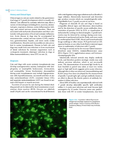
cyclosporin treatment, distemper infection in dogs or (IFA) (Figure 110.1), agglutination assays, and immuno-
feline immunodeficiency virus (FIV) in cats [4].
chromatographic kits.
Subclinically infected animals may display antibody
Diagnosis levels, and therefore positive serologic results may only
indicate previous infection, which is not necessarily
Cats and dogs with acute systemic toxoplasmosis may acute. The demonstration of increased IgG levels by at
develop nonregenerative anemia, leukopenia with lym- least fourfold in paired sera taken at least two weeks
phopenia, or neutrophilic leukocytosis, monocytosis, apart or high IgM levels suggests active acute infection,
and eosinophilia. Serum biochemistry abnormalities although this is often not observed. Immunoblotting and
during acute toxoplasmosis may include hypoproteine- ELISA assays have been developed for the measurement
mia with hypoalbuminemia. Increased activities of ala- of specific T. gondii IgM, IgG, and IgA antibody classes in
nine aminotransferase (ALT), creatinine kinase (CK), an effort to discern the kinetic pattern of antibody for-
and aspartate aminotransferase (AST) are found in ani- mation in feline infection.
mals with hepatic or muscle necrosis. About 80% of healthy cats experimentally infected with
Excretion of oocysts by cats during the limited shed- T. gondii develop detectable specific IgM antibodies
ding period can be detected by fecal examination or pol- within 2–4 weeks post infection and most become IgM
ymerase chain reaction (PCR). Oocysts are relatively seronegative by 16 weeks. However, some cats, particu-
small (10 μM) and can be concentrated by fecal flotation larly those with FIV or ocular toxoplasmosis, may have
Figure 110.1 A positive indirect fluorescent
antigen test serologic response to Toxoplasma
gondii antigen. Source: Courtesy of Dr Monica
Leszkowicz Mazuz, Kimron Veterinary Institute,
Israel.