
Page 1061 - Clinical Small Animal Internal Medicine
P. 1061
109 Fungal Infections 999
History and Clinical Signs Therapy
VetBooks.ir In dogs and cats, Sporothrix spp. most often cause focal The treatment of choice for most dogs and cats with
sporotrichosis is itraconazole. Alternative antifungal
or multifocal cutaneous lesions. These are crusted,
thickened or nodular lesions that often ulcerate or drain drugs for refractory disease include supersaturated
serosanguinous fluid. Most cats have multiple cutaneous potassium or sodium iodide, terbinafine, or ampho
lesions. These lesions may develop as a result of autoin tericin B. Unfortunately, potassium iodide formulations
oculation during grooming or they may be a manifesta often lead to adverse gastrointestinal signs in cats.
tion of a disseminated infection. Localized hyperthermia has been used as adjunct ther
Transmission by inhalation is thought to occur in some apy to treat fixed cutaneous lesions, because most strains
cats that develop pulmonary and nasal cavity disease. of Sporothrix survive poorly at high temperatures. In this
Respiratory signs include sneezing, cough, tachypnea, case, a thermal bag at a temperature of 40–42 °C is
increased respiratory effort, stertor, and/or nasal dis applied at least twice daily to lesions for 15 minutes.
charge. Cats also can develop generalized disease, with
involvement of the lungs, liver, spleen, kidneys, lymph
nodes, and/or testicles. Clinical signs of systemic involve Prognosis
ment include fever, generalized lymphadenomegaly, In general, the prognosis for cure of sporotrichosis is
anorexia, vomiting, and weight loss. Osteoarticular good. Treatment durations of at least 4–6 months are
sporotrichosis has rarely been described in dogs. generally required, but some cats require more than a
year of treatment. Treatment should be continued for a
Diagnosis month after complete resolution of lesions.
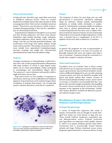
Cytologic examination or histopathology of affected tis
sues from cats reveals pyogranulomatous inflammation Public Health Implications
with large numbers of round to cigar‐shaped yeasts, Inoculation from cat scratches, bites or direct contact
which can be found intracellularly within neutrophils with infected cats are important means by which people
or macrophages and extracellularly (Figure 109.7). acquire sporotrichosis. Sporotrichosis should be consid
The yeasts are 4–6 μm in diameter and typically exhibit a ered as a differential diagnosis for any cat with a draining
single bud with a narrow base. cutaneous lesion, and such cats should be handled with
Dogs tend to have very low numbers of organisms in gloves and hands washed after contact. Owners of
skin lesions, which may not be readily detected. Definitive infected animals should minimize contact, and if contact
diagnosis of sporotrichosis can be made by fungal culture is necessary, it should be recommended that they wear
of aspirates or biopsies from lesions. Use of molecular gloves and wash their hands immediately after handling
assays to identify Sporothrix could also be considered.
the animal. Owners should also be warned of the risk of
exposure to the organism in the environment. Needle‐
stick injuries should be avoided and laboratory person
nel warned of suspect cases.
Miscellaneous Fungus‐Like Infections
(Pythiosis and Rhinosporidiosis)
Etiology/Pathophysiology
A variety of fungus‐like organisms that reside in
aquatic environments and are thought to invade dam
aged host tissues can cause disease in dogs and cats.
These include Pythium insidiosum, Lagenidium spp.
and Rhinosporidium seeberi. Pythium insidiosum
and Lagenidium are oomycetes (water molds), whereas
R. seeberi is an aquatic protistan parasite (class Meso
mycetozoea) that is taxonomically located between
Figure 109.7 Cytology of an impression smear of a skin lesion
from a cat with cutaneous sporotrichosis. Large numbers of animals and fungi. Pythium insidiosum causes severe,
intracellular pleomorphic yeasts are present. progressive gastrointestinal or cutaneous disease in dogs