
Page 1156 - Clinical Small Animal Internal Medicine
P. 1156
1094 Section 10 Renal and Genitourinary Disease
Cytology of tissue acquired by fine needle aspirates has (particularly Masson’s trichrome stain) can aid in assess-
VetBooks.ir limited utility in cases of AKI, but can aid in detection of ment of the severity of fibrosis and provide insight into
the potential for renal recovery. The risk of significant
an infiltrative etiology. Cytology for the diagnosis of
renal lymphosarcoma may produce false‐negative
uremia is severe and platelet dysfunction is present.
results. Therefore, for cases in which lymphosarcoma is hemorrhage secondary to renal biopsy is high when
suspected, histopathologic assessment of tissue may be Ethylene glycol intoxication is an emergency situation
necessary to rule out this etiology. Diagnosis of amyloi- requiring immediate, specific therapy, which makes
dosis and feline infectious peritonitis requires special accurate and timely diagnosis crucial. Commercially
cytologic techniques (e.g., Congo red staining or corona- available in‐house test kits are available.
virus immunocytochemistry, respectively), and these
diagnostic techniques have not been rigorously assessed.
The risk of bleeding secondary to fine needle aspiration Therapy
of the kidneys is low but possible, especially when plate-
let dysfunction is present. Treatment of AKI is primarily aimed at addressing the
Histopathologic samples can be obtained by percuta- underlying cause (if it can be identified and treated) and
neous, ultrasonographically guided needle biopsy, lapa- supportive measures to minimize the clinical sequelae of
roscopy, or surgical wedge biopsy. Histopathology may uremia. This section provides treatment recommenda-
confirm a suspected etiology (e.g., ethylene glycol intoxi- tions for cases of severe AKI, in which severe uremic
cation, renal lymphosarcoma) or it may disclose nonspe- manifestations and abnormalities of acid–base, electro-
cific findings. When AKI cannot be clinically distinguished lyte, and fluid balance dominate the clinical picture.
from end‐stage chronic kidney disease, histopathology Specific doses for many of the drugs discussed are listed
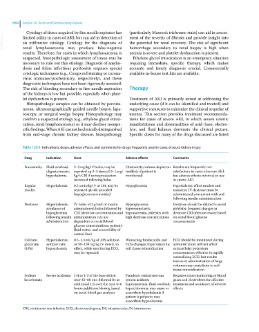
Table 120.4 Indications, doses, adverse effects, and comments for drugs frequently used in cases of acute kidney injury
Drug Indication Dose Adverse effects Comments
Furosemide Fluid overload, 2–5 mg/kg IV bolus, may be Ototoxicity; volume depletion Results are frequently not
oliguria/anuria, repeated up 3–5 times; 0.5–1 mg/ (unlikely if patient is satisfactory in cases of severe AKI,
hyperkalemia kg/h CRI if urine production monitored) but adverse effects minimal so use
increased following bolus in anuric AKI
Regular Hyperkalemia 0.5 units/kg IV or IM, may be Hypoglycemia Hypokalemic effect modest and
insulin repeated q4–6h provided transient; IV dextrose must be
hypoglycemia is avoided administered concurrent with and
following insulin administration
Dextrose Hyperkalemia; IV bolus of 2 g/unit of insulin Hyperglycemia, Dextrose should be diluted to avoid
avoidance of administered; bolus followed by hyperosmolarity, phlebitis; frequent changes in
hypoglycemia CRI (dextrose concentration and hyponatremia, phlebitis with dextrose CRI often necessary based
following insulin administration rate are high dextrose concentrations on serial blood glucose
administration dependent on serial blood measurements
glucose concentrations, patient’s
fluid status, and accessibility of
central line)
Calcium Hyperkalemia; 0.5–1.5 mL/kg of 10% solution Worsening bradycardia and ECG should be monitored during
gluconate symptomatic or 50–150 mg/kg IV slowly, to ECG changes; hypercalcemia; administration; will not affect
(10%) hypocalcemia effect, while monitoring ECG, soft tissue mineralization extracellular potassium
may be repeated concentration; effective in rapidly
normalizing ECG, but results
transient; administration of large
volumes may contribute to soft
tissue mineralization
Sodium Severe acidemia 1/4 to 1/3 of the base deficit Paradoxic central nervous Requires close monitoring of blood
bicarbonate over 30–60 min followed by an system acidosis; gases and electrolytes for effective
additional 1/4 over the next 4–6 hypernatremia; fluid overload; treatment and avoidance of adverse
hours; additional dosing based hypochloremia; may cause or effects
on serial blood gas analyses exacerbate hypokalemia if
patient is polyuric; may
exacerbate hypocalcemia
CRI, continuous rate infusion; ECG, electrocardiogram; IM, intramuscular; IV, intravenous.