
Page 1220 - Clinical Small Animal Internal Medicine
P. 1220
1158 Section 10 Renal and Genitourinary Disease
prostate tends to feel enlarged but it is smooth, symmet-
VetBooks.ir ric, and rarely painful. The median raphe may not be as
obvious to palpate. The enlargement may cause the pros-
tate to move cranially into the abdominal cavity, and
therefore can be difficult to palpate rectally without the
help of concurrent abdominal palpation. Prostatic cysts
can rarely be identified. Rectal palpation should also
include the dorsal portion of the distal colon to feel for
enlarged iliac lymph nodes. With BPH, lymphadenopa-
thy should not be present. Constipation may be possible,
but the rest of the physical exam is usually within
normal limits.
Complete blood count (CBC) and chemistry panel are
unremarkable in dogs with BPH and noninfected pros-
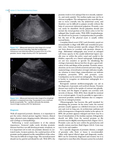
Figure 124.1 Ultrasound transverse view image of a normal tatic cysts. Human prostate‐specific antigen (PSA) has
prostate of an intact male dog. Note the homogenous not been shown to correlate with prostatic disease in
echotexture and relatively uniform but prominent size of the dogs. Abdominal radiographs may reveal an enlarged
prostate. Source: Image courtesy of Dr Tim Spotswood. soft tissue opacity in the caudal abdominal region usu-
ally near or within the pelvic canal and caudal to the
bladder, especially on a lateral radiograph. Radiographs
are not very sensitive or specific for identifying the
etiology of prostatic disease but they do give a good indi-
cation of size and shape of the prostate. Prostatic miner-
alization can be seen in both neutered and intact dogs. In
neutered dogs, mineralization strongly indicates neopla-
sia, whereas in intact dogs, mineralization can indicate
neoplasia, prostatitis, BPH, and prostatic cysts.
Constipation can be noted on radiographs. The prostate
is harder to recognize on abdominal radiographs in a
neutered male.
Retrograde contrast urethrocystography can aid in
the diagnosis of prostatic and urethral disease. Prostatic
disease may result in the uptake of contrast into glandu-
lar tissue, and the degree of uptake can correlate with
severity of disease. With BPH, there should be minimal
to no contrast uptake. It may be possible to appreciate a
Figure 124.2 Ultrasound transverse view image of an atrophied narrowed prostatic urethral diameter due to compres-
prostate in a neutered male dog. The prostate is very small and sion by an enlarged prostate.
barely recognizable. The + symbols delineate the prostate. Ultrasonography has become the gold standard for
Source: Image courtesy of Dr Tim Spotswood. visualizing the prostate. In the intact male, the normal
prostate usually appears as a bilobed structure on short
axis and oval on long axis, its architecture uniform and
Diagnosis
with a homogenous echogenicity similar to the spleen. In
With all suspected prostatic diseases, it is important to BPH, the prostate appears enlarged and there may be a
put the entire clinical picture together: history, clinical loss of visualization of the median septum. Echogenicity
signs, physical exam, imaging results, laboratory results, should not differ from the normal prostate or the
and cytology or histopathology. prostate may be slightly more hyperechoic (Figure 124.3).
Performing a rectal examination is of the utmost Prostatic cysts can be seen within the parenchyma and
importance. Neutered males rarely develop prostatic usually contain anechoic fluid (Figure 124.4). Cysts may
diseases but they can still develop prostatic neoplasia, so vary in size and number.
it is important not to rule out prostatic diseases in cas- For a specific diagnosis of prostatic disease, a sample
trated males. In most patients, the caudal portion of the of prostatic cells, fluid, or tissue is recommended.
prostate can be palpated on the ventral floor of the colon. Often with typical clinical signs and diagnostic results,
This may be difficult in larger dogs. The normal prostate BPH can be tentatively diagnosed and treated, but it is
should not be painful and should be soft. With BPH, the important to note that other diseases cannot be excluded.