Page 1572 - Clinical Small Animal Internal Medicine
P. 1572

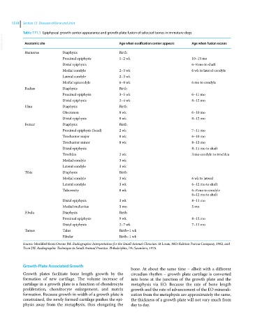
1510  Section 13  Diseases of Bone and Joint

            Table 171.1  Epiphyseal growth center appearance and growth plate fusion of selected bones in immature dogs
  VetBooks.ir  Anatomic site                             Age when ossification center appears  Age when fusion occurs


             Humerus          Diaphysis                  Birth
                              Proximal epiphysis         1–2 wk                            10–13 mo
                              Distal epiphysis                                             6–8 mo to shaft
                              Medial condyle             2–3 wk                            6 wk to lateral condyle
                              Lateral condyle            2–3 wk
                              Medial epicondyle          6–8 wk                            6 mo to condyle
             Radius           Diaphysis                  Birth
                              Proximal epiphysis         3–5 wk                            6–11 mo
                              Distal epiphysis           2–4 wk                            8–12 mo
             Ulna             Diaphysis                  Birth
                              Olecranon                  8 wk                              6–10 mo
                              Distal epiphysis           8 wk                              8–12 mo
             Femur            Diaphysis                  Birth
                              Proximal epiphysis (head)  2 wk                              7–11 mo
                              Trochanter major           8 wk                              6–10 mo
                              Trochanter minor           8 wk                              8–13 mo
                              Distal epiphysis                                             8–11 mo to shaft
                              Trochlea                   2 wk                              3 mo condyle to trochlea
                              Medial condyle             3 wk
                              Lateral condyle            3 wk
             Tibia            Diaphysis                  Birth
                              Medial condyle             3 wk                              6 wk to lateral
                              Lateral condyle            3 wk                              6–12 mo to shaft
                              Tuberosity                 8 wk                              6–8 mo to condyle
                                                                                           6–12 mo to shaft
                              Distal epiphysis           3 wk                              8–11 mo
                              Medial malleolus           3 mo                              5 mo
             Fibula           Diaphysis                  Birth
                              Proximal epiphysis         9 wk                              8–12 mo
                              Distal epiphysis           2–7 wk                            7–11 mo
             Tarsus           Talus                      Birth–1 wk
                              Fibular                    Birth–1 wk

            Source: Modified from Owens JM. Radiographic Interpretation for the Small Animal Clinician. St Louis, MO: Ralston Purina Company, 1982, and
            Ticer JW. Radiographic Technique in Small Animal Practice. Philadelphia, PA: Saunders, 1975.


            Growth‐Plate Associated Growth
                                                              bone. At about the same time – albeit with a different
            Growth plates facilitate bone length growth by the   circadian rhythm – growth plate cartilage is converted
              formation  of  new  cartilage.  The  volume  increase  of   into bone at the junction of the growth plate and the
              cartilage in a growth plate is a function of chondrocyte   metaphysis via EO.  Because the  rate of  bone  length
            proliferation, chondrocyte enlargement, and matrix   growth and the rate of advancement of the EO minerali-
              formation. Because growth in width of a growth plate is   zation from the metaphysis are approximately the same,
            constrained, the newly formed cartilage pushes the epi-  the thickness of a growth plate will not vary much from
            physis  away  from  the  metaphysis,  thus  elongating  the   day to day.
   1567   1568   1569   1570   1571   1572   1573   1574   1575   1576   1577