
Page 600 - Clinical Small Animal Internal Medicine
P. 600
568 Section 6 Gastrointestinal Disease
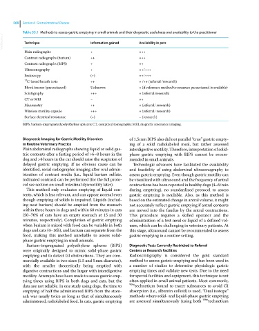
Table 53.1 Methods to assess gastric emptying in small animals and their diagnostic usefulness and availability to the practitioner
VetBooks.ir Technique Information gained Availability in pets
Plain radiographs + +++
Contrast radiographs (barium) ++ +++
Contrast radiographs (BIPS) + ++
Ultrasonography + ++/+++
Endoscopy (+) ++/+++
13 C‐based breath tests ++ + /++ (referral /research)
Blood tracers (paracetamol) Unknown + (if reference method to measure paracetamol is available)
Scintigraphy +++ + (referral/research)
CT or MRI − ++
Manometry ++ + (referral/ research)
Wireless motility capsule +++ + (referral/ research)
Surface electrical resistance (+) − (research)
BIPS, barium‐impregnated polyethylene spheres; CT, computed tomography; MRI, magnetic resonance imaging.
Diagnostic Imaging for Gastric Motility Disorders of 1.5 mm BIPS also did not parallel “true” gastric empty-
in Routine Veterinary Practice ing of a solid radiolabeled meal, but rather assessed
Plain abdominal radiographs showing liquid or solid gas- interdigestive motility. Therefore, interpretation of solid‐
tric contents after a fasting period of >6–8 hours in the phase gastric emptying with BIPS cannot be recom-
dog and >4 hours in the cat should raise the suspicion of mended in small animals.
delayed gastric emptying. If no obvious cause can be Technologic advances have facilitated the availability
identified, serial radiographic imaging after oral admin- and feasibility of using abdominal ultrasonography to
istration of contrast media (i.e., liquid barium sulfate, assess gastric emptying. Even though gastric motility can
iodinated contrast) can be performed (for the full proto- be visualized with ultrasound and the frequency of antral
col see section on small intestinal dysmotility later). contractions has been reported in healthy dogs (4–6/min
This method only evaluates emptying of liquid con- during emptying), no standardized protocol to assess
tents, which is less relevant, and can appear normal even gastric emptying is available. Also, as this method is
though emptying of solids is impaired. Liquids (includ- based on the estimated change in antral volume, it might
ing neat barium) should be emptied from the stomach not accurately reflect gastric emptying if antral contents
within three hours in dogs and within 60 minutes in cats are moved into the fundus by the antral contractions.
(50–70% of cats have an empty stomach at 15 and 30 This procedure requires a skilled operator and the
minutes, respectively). Completion of gastric emptying administration of a test meal or liquid of a defined vol-
when barium is mixed with food can be variable in both ume, which can be challenging in veterinary patients. At
dogs and cats (4–16h), and barium can separate from the this stage, ultrasound cannot be recommended to assess
food, making this method unreliable to assess solid‐ gastric emptying in a routine setting.
phase gastric emptying in small animals.
Barium‐impregnated polyethylene spheres (BIPS) Diagnostic Tests Currently Restricted to Referral
were originally designed to mimic solid‐phase gastric Centers or Research Facilities
emptying and to detect GI obstructions. They are com- Radioscintigraphy is considered the gold standard
mercially available in two sizes (1.5 and 5 mm diameter), method to assess gastric emptying and has been used in
with the smaller theoretically being emptied with a number of studies to determine physiologic gastric
digestive contractions and the larger with interdigestive emptying times and validate new tests. Due to the need
motility. Attempts have been made to assess gastric emp- for special facilities and equipment, this technique is not
tying times using BIPS in both dogs and cats, but the often applied in small animal patients. Most commonly,
data are not reliable. In one study using dogs, the time to 99m technetium bound to tracer substances to avoid GI
emptying of half the administered BIPS from the stom- absorption (i.e., albumin colloid) is used. “Dual isotope”
ach was nearly twice as long as that of simultaneously methods where solid‐ and liquid‐phase gastric emptying
administered, radiolabeled food. In cats, gastric emptying are assessed simultaneously (using both 99m technetium