
Page 605 - Clinical Small Animal Internal Medicine
P. 605
53 Motility Disorders of the Alimentary Tract 573
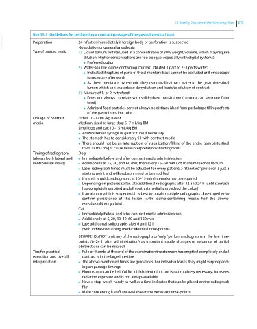
VetBooks.ir Box 53.1 Guidelines for performing a contrast passage of the gastrointestinal tract
Preparation
24 h fast or immediately if foreign body or perforation is suspected
Type of contrast media No sedation or general anesthesia
1) Liquid barium sulfate (used at a concentration of 30% weight/volume, which may require
dilution. Higher concentrations are too opaque, especially with digital systems)
Preferred option
●
2) Water‐soluble iodine‐containing contrast (diluted 1 part to 2–3 parts water)
Indicated if rupture of parts of the alimentary tract cannot be excluded or if endoscopy
●
is necessary afterwards
As these media are hypertonic, they osmotically attract water to the gastrointestinal
●
lumen which can exacerbate dehydration and leads to dilution of contrast
3) Mixture of 1. or 2. with food
Does not always correlate with solid‐phase transit time (contrast can separate from
●
food)
Admixed food particles cannot always be distinguished from pathologic filling defects
●
of the gastrointestinal tube
Dosage of contrast Either 10–12 mL/kg BM or
media Medium‐sized to large dog: 5–7 mL/kg BM
Small dog and cat: 10–15 mL/kg BM
Administer via syringe or gastric tube if necessary
●
The stomach has to considerably fill with contrast media
●
There should not be an interruption of visualization/filling of the entire gastrointestinal
●
tract, as this might cause false interpretation of radiographs
Timing of radiographs Dog
(always both lateral and ● Immediately before and after contrast media administration
ventrodorsal views) ● Additionally at 15, 30, and 60 min; then every 15–60 min until barium reaches rectum
Later radiograph times must be adjusted for every patient; a “standard” protocol is just a
●
starting point and will probably need to be modified
If transit is quick, radiographs at 10–15‐min intervals may be required
●
Depending on pictures so far, late additional radiographs after 12 and 24 h (until stomach
●
has completely emptied and all contrast media has reached the colon)
If an abnormalitiy is suspected, it is best to obtain multiple radiographs close together to
●
confirm persistence of the lesion (with iodine‐containing media: half the above‐
mentioned time‐points)
Cat
Immediately before and after contrast media administration
●
Additionally at 5, 20, 30, 40, 60 and 120 min
●
Late additional radiographs after 6 and 12 h
●
(with iodine‐containing media: identical time‐points)
BEWARE: Do NOT omit any of the radiographs or “only” perform radiographs at the late time‐
points (6–24 h after administration) as important subtle changes or evidence of partial
obstructions can be missed!
Tips for practical ● Rule of thumb: at the end of the examination the stomach has emptied completely and all
execution and overall contrast is in the large intestine
interpretation ● The above‐mentioned times are guidelines. For individual cases they might vary depend-
ing on passage timings
Fluoroscopy can be helpful for initial orientation, but is not routinely necessary, increases
●
radiation exposure and is not always available
Have a stop‐watch handy as well as a time indicator that can be placed on the radiograph
●
film
Make sure enough staff are available at the necessary time‐points
●