
Page 643 - Clinical Small Animal Internal Medicine
P. 643
57 Rectoanal Diseases – Medical and Surgical Management 611
prevent fecal incontinence; and ligate the anal sac duct imperforate anus), type III (as with type II but the rectum
VetBooks.ir near its opening, removing it en bloc with the gland itself. ends further cranially), and type IV (terminal rectum and
anus may be normal but there is a discontinuity in the
These goals are most readily achieved with the patient
positioned in sternal recumbency in a perineal stand
These animals appear normal until 2–4 weeks of age at
with the tail in an elevated position and an anal purse‐ rectum with a blind pouch within the pelvic canal).
string suture placed cranial to the opening of the anal sac which time they become unthrify, anorexic, and restless
ducts. If a closed technique is chosen, the anal sac itself with abdominal distension, possibly with perineal
can be distended with various materials (e.g., yarn, sili- bulging.
cone sealant, Foley balloon catheter, plaster of Paris, agar Rectoanal strictures appear to be more common in
base gel, dental molds, etc.) to facilitate palpation of cats than dogs, but can occur in either species, most typ-
the gland itself during dissection. Closure is typically ically after trauma (iatrogenic or spontaneous). Clinical
performed in two layers; 3‐0 or 4‐0 monofilament signs may vary depending on the underlying cause, but
absorbable suture for the subcutaneous tissues and include tenesmus, dyschezia, hematochezia, and passing
either similarly sized nylon skin sutures or an intrader- of ribbon‐like feces. Megacolon is a possible sequel,
mal skin closure can be used. especially in cats with chronic partial rectal narrowing
(e.g., due to malunion after pelvic fractures).
Prognosis
The prognosis for patients with anal sac impaction is gen- Pathophysiology
erally excellent, regardless of whether they require surgi- The functional diameter of the distal colon, rectum or
cal intervention or respond to medical management. anus is reduced, due to either congenital (atresia ani) or
acquired anatomic abnormalities.
Atresia Ani and Rectoanal Strictures
Diagnosis and Medical Management
Clinical Presentation Atresia ani may be diagnosed at the time of birth, or in
the neonatal period, with failure of the infant to defecate
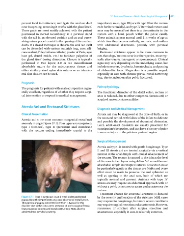
Atresia ani is the most common congential rectal/anal and possibly the development of abdominal distension.
anomaly in dogs (Figure 57.1). Four types are recognized: Later, adult‐onset disorders are usually presented for
type I (stenosis), type II (persistent anal membrane constipation/obstipation, and can have a history of prior
with the rectum ending immediately cranial to the
trauma or injury to the pelvis or perianal region.
Surgical Management
Atresia ani type I is treated with gentle bougienage. Type
II and III atresia ani are treated surgically via a vertical
incision at the anal dimple with caudal advancement of
the rectum. The rectum is sutured to the skin at the level
of the anus in two layers using 4‐0 or 5‐0 monofilament
absorbable simple interrupted sutures. Dissection must
be particularly gentle as the tissues are friable and every
effort must be made to preserve the anal sphincter as
well as opening to the anal sacs, both of which are
typically normal and present. Animals with type IV
atresia ani may require an abdominal approach with or
without a pelvic osteotomy to access and anastomose the
rectum.
Treatment chosen for anorectal strictures is dictated
Figure 57.1 Type II atresia ani in an 8‐week‐old mixed‐breed by the severity and location of the stricture. Mild cases
puppy. Note the imperforate anus and absence of rectal lumen. may respond to bougienage, but more severe conditions
This particular puppy presented later than is typical for this
disorder due to the concurrent presence of a rectovaginal fistula may require surgical resection and anastomosis. However,
that prevented colonic and rectal obstruction. Note also the recurrence of stricture after surgical resection and
abnormalities in vulval anatomy. anastomosis, especially in cats, is relatively common.