
Page 648 - Clinical Small Animal Internal Medicine
P. 648
616 Section 6 Gastrointestinal Disease
deeply sedated or anesthetized for these procedures. In
VetBooks.ir some cases, foreign bodies can be broken up using rec-
tally passed heavy forceps, aiding their eventual passage.
Surgical Management
Surgical management of rectal foreign bodies is not typi-
cally required as nonsurgical removal is almost always
possible. In the event of rectal tears or trauma develop-
ing as a consequence of a rectal foreign body, surgical
repair of the defects may be indicated (see Rectal Tears
and Trauma section).
Prognosis
Most rectal foreign bodies are removed with minimal to
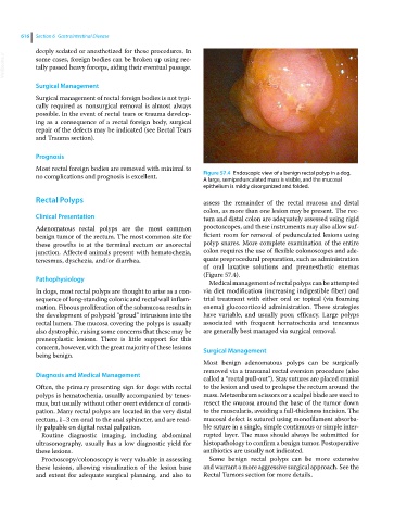
no complications and prognosis is excellent. Figure 57.4 Endoscopic view of a benign rectal polyp in a dog.
A large, semipedunculated mass is visible, and the mucosal
epithelium is mildly disorganized and folded.
Rectal Polyps assess the remainder of the rectal mucosa and distal
colon, as more than one lesion may be present. The rec-
Clinical Presentation tum and distal colon are adequately assessed using rigid
Adenomatous rectal polyps are the most common proctoscopes, and these instruments may also allow suf-
benign tumor of the rectum. The most common site for ficient room for removal of pedunculated lesions using
these growths is at the terminal rectum or anorectal polyp snares. More complete examination of the entire
junction. Affected animals present with hematochezia, colon requires the use of flexible colonoscopes and ade-
tenesmus, dyschezia, and/or diarrhea. quate preprocedural preparation, such as administration
of oral laxative solutions and preanesthetic enemas
(Figure 57.4).
Pathophysiology
Medical management of rectal polyps can be attempted
In dogs, most rectal polyps are thought to arise as a con- via diet modification (increasing indigestible fiber) and
sequence of long‐standing colonic and rectal wall inflam- trial treatment with either oral or topical (via foaming
mation. Fibrous proliferation of the submucosa results in enema) glucocorticoid administration. These strategies
the development of polypoid “proud” intrusions into the have variable, and usually poor, efficacy. Large polyps
rectal lumen. The mucosa covering the polyps is usually associated with frequent hematochezia and tenesmus
also dystrophic, raising some concerns that these may be are generally best managed via surgical removal.
preneoplastic lesions. There is little support for this
concern, however, with the great majority of these lesions Surgical Management
being benign.
Most benign adenomatous polyps can be surgically
removed via a transanal rectal eversion procedure (also
Diagnosis and Medical Management
called a “rectal pull‐out”). Stay sutures are placed cranial
Often, the primary presenting sign for dogs with rectal to the lesion and used to prolapse the rectum around the
polyps is hematochezia, usually accompanied by tenes- mass. Metzenbaum scissors or a scalpel blade are used to
mus, but usually without other overt evidence of consti- resect the mucosa around the base of the tumor down
pation. Many rectal polyps are located in the very distal to the muscularis, avoiding a full‐thickness incision. The
rectum, 1–3 cm orad to the anal sphincter, and are read- mucosal defect is sutured using monofilament absorba-
ily palpable on digital rectal palpation. ble suture in a single, simple continuous or simple inter-
Routine diagnostic imaging, including abdominal rupted layer. The mass should always be submitted for
ultrasonography, usually has a low diagnostic yield for histopathology to confirm a benign tumor. Postoperative
these lesions. antibiotics are usually not indicated.
Proctoscopy/colonoscopy is very valuable in assessing Some benign rectal polyps can be more extensive
these lesions, allowing visualization of the lesion base and warrant a more aggressive surgical approach. See the
and extent for adequate surgical planning, and also to Rectal Tumors section for more details.