
Page 649 - Clinical Small Animal Internal Medicine
P. 649
57 Rectoanal Diseases – Medical and Surgical Management 617
Prognosis
VetBooks.ir The prognosis for dogs with benign rectal polyps is good,
but a regular monitoring schedule should be instituted
to assess for recurrence and potentially progression of
disease towards a malignant state. If caught early in
the course of disease, lesions undergoing malignant
transformation treated appropriately (e.g., carcinoma in
situ resected with histologically complete surgical mar-
gins) can be successfully treated with good long‐term
survival.
Rectal Prolapse
Clinical Presentation
Depending on whether only anal mucosa is protruding
or all layers of the terminal rectum are protruding as a
cylindrical mass, rectal prolapse can be classified as
either partial or complete, respectively. Most commonly
occurring in younger animals, prolapse is typically a sec-
ondary condition, caused by tenesmus from presence of
parasites, foreign bodies, or urogenital diseases.
Pathophysiology
Rectal prolapse is generally due to excessive straining,
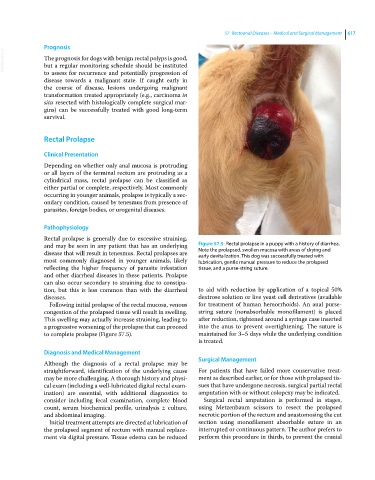
and may be seen in any patient that has an underlying Figure 57.5 Rectal prolapse in a puppy with a history of diarrhea.
disease that will result in tenesmus. Rectal prolapses are Note the prolapsed, swollen mucosa with areas of drying and
early devitalization. This dog was successfully treated with
most commonly diagnosed in younger animals, likely lubrication, gentle manual pressure to reduce the prolapsed
reflecting the higher frequency of parasite infestation tissue, and a purse‐string suture.
and other diarrheal diseases in these patients. Prolapse
can also occur secondary to straining due to constipa-
tion, but this is less common than with the diarrheal to aid with reduction by application of a topical 50%
diseases. dextrose solution or live yeast cell derivatives (available
Following initial prolapse of the rectal mucosa, venous for treatment of human hemorrhoids). An anal purse‐
congestion of the prolapsed tissue will result in swelling. string suture (nonabsorbable monofilament) is placed
This swelling may actually increase straining, leading to after reduction, tightened around a syringe case inserted
a progressive worsening of the prolapse that can proceed into the anus to prevent overtightening. The suture is
to complete prolapse (Figure 57.5). maintained for 3–5 days while the underlying condition
is treated.
Diagnosis and Medical Management
Surgical Management
Although the diagnosis of a rectal prolapse may be
straightforward, identification of the underlying cause For patients that have failed more conservative treat-
may be more challenging. A thorough history and physi- ment as described earlier, or for those with prolapsed tis-
cal exam (including a well‐lubricated digital rectal exam- sues that have undergone necrosis, surgical partial rectal
ination) are essential, with additional diagnostics to amputation with or without colopexy may be indicated.
consider including fecal examination, complete blood Surgical rectal amputation is performed in stages,
count, serum biochemical profile, urinalysis ± culture, using Metzenbaum scissors to resect the prolapsed
and abdominal imaging. necrotic portion of the rectum and anastomosing the cut
Initial treatment attempts are directed at lubrication of section using monofilament absorbable suture in an
the prolapsed segment of rectum with manual replace- interrupted or continuous pattern. The author prefers to
ment via digital pressure. Tissue edema can be reduced perform this procedure in thirds, to prevent the cranial