
Page 651 - Clinical Small Animal Internal Medicine
P. 651
57 Rectoanal Diseases – Medical and Surgical Management 619
The clinical signs accompanying rectal tumors are Surgical Management
VetBooks.ir similar to most other rectal diseases, featuring histories In preparation for surgery of the rectum, many surgeons
of straining to defecate and occasionally the presence of
recommend feeding a low‐residue diet for 4–5 days
narrow “ribbon‐like” stool due to partial restriction of
the rectal lumen. Hematochezia is variably present, preoperatively and avoiding enemas in the three days
preceding surgery. Perioperative antibiotic cover typi-
depending on the involvement of the mucosa. Tumors cally consists of cephalosporins but some surgeons
such as adenocarcinoma and mucosal lymphoma often prefer metronidazole or other agents that may target
present with histories of hematochezia, while tumors colorectal anaerobic bacteria. The need for postopera-
affecting the muscularis and deeper mural layers, such as tive antibiotics depends on degree of contamination
leiomyoma or GI stromal tumors, usually do not feature during surgery, but is typically not necessary beyond
hematochezia.
24 hours postoperatively.
Rectal surgery can be divided into two general catego-
ries: procedures that can be performed from a perineal
Pathophysiology
or transanal approach and procedures that require an
Rectal tumors result from neoplastic or hyperplastic intraabdominal approach. The first is the simpler and
changes in normal tissue of the rectum or anal sphinc- more commonly performed option. The exact distance
ter. Any of the various tissue types within the rectum accessible depends on the patient’s size and should be
can be involved (mucosa, secretory epithelia, smooth assessed via digital rectal exam prior to deciding upon
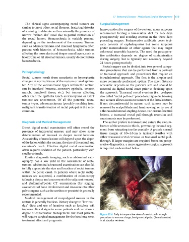
muscle, lymphoid tissue, etc.), but tumors affecting the approach. Transanal rectal eversion (i.e., prolapse;
other than the epithelia (such as leiomyoma or GIST also called “rectal pull‐out” procedure; Figure 57.6) using
tumors) are uncommon. Of all the potential rectal stay sutures allows access to tumors of the distal rectum.
tumor types, adenocarcinoma (possibly resulting from If not circumferential in nature, such tumors may be
malignant transformation of rectal polyps) is the most removed by scalpel blade and hand‐sewing, or by use of
common. a thoracoabdominal stapling device. For circumferential
lesions, a transanal rectal pull‐through resection and
anastomosis may be performed.
Diagnosis and Medical Management The author prefers to transect and suture the circum-
ference of the rectum in thirds, preventing the orad seg-
Direct digital rectal examination will often reveal the ment from retracting too far cranially. A grossly normal
presence of intrarectal masses, and may allow some tissue margin of 0.5–1.0 cm is typically feasible with
determination of mucosal vs deeper mural location. either transanal rectal eversion or transanal rectal pull‐
Accessibility of mass lesions will depend upon the depth through. If larger margins are required based on preop-
of the lesion within the rectum, the size of the animal and erative diagnostics, a more aggressive surgical approach
examiner’s reach. Effective digital rectal examination is required, as described below.
often requires sedation of the patient, particularly with
smaller animals.
Routine diagnostic imaging, such as abdominal radi-
ography, has a low yield in the assessment of rectal
tumors. Abdominal ultrasound examination can also fail
to fully appreciate the size and location of rectal tumors
within the pelvic canal. In patients where rectal malig-
nancies are suspected, a combination of colonoscopy
(allowing biopsy and assessment of the adjacent mucosa)
and abdominal/pelvic CT examination (for staging,
assessment of bone involvement and invasion into other
pelvic organs such as the urethra or prostate) is generally
recommended.
Medical management of nonpolypoid masses in the
rectum is generally fruitless. Dietary change to “low‐resi-
due” diets and use of laxatives such as lactulose will
improve clinical signs in some patients and can allow a
degree of conservative management, but most patients Figure 57.6 Early intraoperative view of a rectal pull‐through
will require surgical management for the best long‐term procedure to remove a large, benign rectal polyp (2 cm diameter)
treatment effect and prognosis. in a Labrador retriever.