
Page 674 - Clinical Small Animal Internal Medicine
P. 674
642 Section 7 Diseases of the Liver, Gallbladder, and Bile Ducts
fluid and inorganic solutes. The bile ducts contribute
VetBooks.ir substantially to this bile formation and flow, with secre-
tion of bicarbonate, chloride, and water. Approximately
50% of bile is released immediately into the gastrointesti-
nal tract, while the remainder is stored and concentrated
in the gallbladder. Intrahepatic cholestasis disrupts this
bile flow mainly at the level of the hepatocytes, their can-
aliculi or the bile ductules, in the periportal zone (zone 1)
of the liver lobule, whereas extrahepatic cholestasis
results from obstruction of the common bile duct. In
both of these situations, significant hepatic reserve and
compensatory changes have the net result that, typically,
overt clinical signs of cholestasis, such as icterus (jaun-
dice), only result from diffuse liver pathology or severe
impairment of the common bile duct flow.
Intrahepatic cholestasis occurs commonly to varying
degrees in most clinical cases, with various proposed
mechanisms involved. Damage to tight junctions sepa-
rating the bile canaliculi from blood sinusoids can result
from endotoxemia, sepsis, some Leptospira infections or
following an adverse drug reaction. At the cellular level,
swollen hepatocytes may directly obstruct bile flow
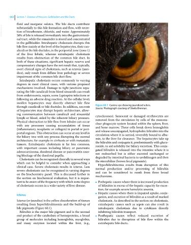
through canaliculi or bile ductules. In addition, necrosis Figure 60.1 Canine eye showing jaundiced sclera.
Source: Photograph courtesy of Sheila Brennan.
of hepatocytes may disrupt hepatic architecture, allow-
ing communication between canaliculi and sinusoidal cytochromes). Senescent or damaged erythrocytes are
lymph or blood, aided by the inherent biliary pressure. removed from the circulation by cells of the mononu-
Physical obstruction to bile flow from lobules can occur clear phagocyte system located within the spleen, liver,
with any processes causing accumulation of tissue and bone marrow. These cells break down hemoglobin
(inflammatory, neoplastic or collagen) in portal or peri- and release unconjugated, hydrophobic bilirubin into the
portal regions. This obstruction can occur at any level in circulation where it is carried, reversibly bound to albu-
the biliary tree with any process disrupting the normal min, to the liver for clearance. The hepatocytes take up
architecture, for example in cirrhosis or with metastatic the bilirubin and conjugate it, predominantly with glucu-
tumors. Extrahepatic cholestasis is far less common, ronide, to aid solubility for biliary excretion. This conju-
with important causes including biliary or pancreatic gated bilirubin is released into the intestine where it is
adenocarcinoma, duodenal disease or pancreatitis caus- not reabsorbed but is either excreted unchanged or
ing blockage of the duodenal papilla. degraded by intestinal bacteria to urobilinogen and then
Cholestasis can be recognized clinically in several ways
which can be helpful to consider when approaching a the stercobilins (brown fecal pigments).
Hyperbilirubinemia results from disruption of this
clinical case. Severe cholestasis results in icterus. Less normal production and/or processing of bilirubin
severe cholestasis can be recognized in varying degrees and can be considered to result from three broad
on the biochemistry panel. This is discussed further in mechanisms.
the section on biochemical evaluation, but it is impor-
tant to be aware of the frequency with which some degree ● Prehepatic causes where there is increased production
of cholestasis occurs in a wide variety of liver disease. of bilirubin in excess of the hepatic capacity for excre-
tion, for example severe hemolytic anemia.
Hepatic causes where there is impaired uptake, conju-
Icterus ●
gation, and excretion of bilirubin as a result of marked
Icterus (or jaundice) is the yellow discoloration of tissues cholestasis. As described in the section on cholestasis,
resulting from hyperbilirubinemia and the build‐up of extrahepatic causes such as sepsis can also result in
bile pigments (Figure 60.1). intrahepatic cholestasis due to cytokines directly
Bilirubin is the major bile pigment and is the normal inhibiting bilirubin transport.
end‐product of the catabolism of hemoproteins, a broad ● Posthepatic causes reflect reduced excretion of
group of molecules including hemoglobin, myoglobin, bilirubin due to disruption of bile flow within the
and many enzymes located within the liver, (e.g., extrahepatic bile ducts.