Page 678 - Clinical Small Animal Internal Medicine
P. 678
646  Section 7  Diseases of the Liver, Gallbladder, and Bile Ducts


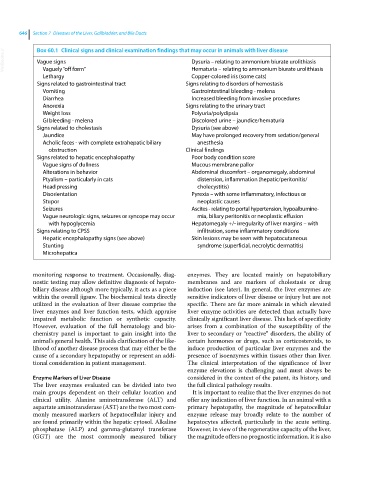
  VetBooks.ir  Box 60.1  Clinical signs and clinical examination findings that may occur in animals with liver disease
             Vague signs
                                                                Dysuria – relating to ammonium biurate urolithiasis
               Vaguely “off form”
               Lethargy                                         Hematuria – relating to ammonium biurate urolithiasis
                                                                Copper‐colored iris (some cats)
             Signs related to gastrointestinal tract          Signs relating to disorders of hemostasis
               Vomiting                                         Gastrointestinal bleeding ‐ melena
               Diarrhea                                         Increased bleeding from invasive procedures
               Anorexia                                       Signs relating to the urinary tract
               Weight loss                                      Polyuria/polydipsia
               GI bleeding ‐ melena                             Discolored urine – jaundice/hematuria
             Signs related to cholestasis                       Dysuria (see above)
               Jaundice                                         May have prolonged recovery from sedation/general
               Acholic feces ‐ with complete extrahepatic biliary   anesthesia
                 obstruction                                  Clinical findings
             Signs related to hepatic encephalopathy            Poor body condition score
               Vague signs of dullness                          Mucous membrane pallor
               Alterations in behavior                          Abdominal discomfort – organomegaly, abdominal
               Ptyalism – particularly in cats                   distension, inflammation (hepatic/peritonitis/
               Head pressing                                     cholecystitis)
               Disorientation                                   Pyrexia – with some inflammatory, infectious or
               Stupor                                            neoplastic causes
               Seizures                                         Ascites ‐ relating to portal hypertension, hypoalbumine-
               Vague neurologic signs, seizures or syncope may occur   mia, biliary peritonitis or neoplastic effusion
                 with hypoglycemia                              Hepatomegaly +/‐ irregularity of liver margins – with
             Signs relating to CPSS                              infiltration, some inflammatory conditions
               Hepatic encephalopathy signs (see above)         Skin lesions may be seen with hepatocutaneous
               Stunting                                          syndrome (superficial, necrolytic dermatitis)
               Microhepatica


              monitoring response to treatment. Occasionally, diag-  enzymes. They are located mainly on hepatobiliary
            nostic testing may allow definitive diagnosis of hepato-  membranes and are markers of cholestasis or drug
            biliary disease although more typically, it acts as a piece   induction (see later). In general, the liver enzymes are
            within the overall jigsaw. The biochemical tests directly   sensitive indicators of liver disease or injury but are not
            utilized in the evaluation of liver disease comprise the   specific. There are far more animals in which elevated
            liver  enzymes  and  liver  function  tests,  which  appraise   liver enzyme activities are detected than actually have
            impaired metabolic function or synthetic capacity.   clinically significant liver disease. This lack of specificity
            However, evaluation of the full hematology and bio-  arises from a combination of the susceptibility of the
            chemistry panel is important to gain insight into the   liver to secondary or “reactive” disorders, the ability of
              animal’s general health. This aids clarification of the like-  certain hormones or drugs, such as corticosteroids, to
            lihood of another disease process that may either be the   induce production of particular liver enzymes and the
            cause of a secondary hepatopathy or represent an addi-  presence of isoenzymes within tissues other than liver.
            tional consideration in patient management.       The  clinical  interpretation  of  the  significance  of  liver
                                                              enzyme elevations is challenging and must always be
            Enzyme Markers of Liver Disease                   considered in the context of the patent, its history, and
            The liver enzymes evaluated can be divided into two   the full clinical pathology results.
            main groups dependent on their cellular location and   It is important to realize that the liver enzymes do not
            clinical utility. Alanine aminotransferase (ALT) and   offer any indication of liver function. In an animal with a
            aspartate aminotransferase (AST) are the two most com-  primary hepatopathy, the magnitude of hepatocellular
            monly measured markers of hepatocellular injury and   enzyme release may broadly relate to the number of
            are found primarily within the hepatic cytosol. Alkaline   hepatocytes affected, particularly in the acute setting.
            phosphatase (ALP) and gamma‐glutamyl  transferase   However, in view of the regenerative capacity of the liver,
            (GGT)  are the  most  commonly  measured  biliary   the magnitude offers no prognostic information. It is also

