
Page 774 - Clinical Small Animal Internal Medicine
P. 774
742 Section 8 Neurologic Disease
Spinal Cord Instability Clinical Assessment of the Spinal Injury Patient
VetBooks.ir consequences for the patient. Once systemic assessment and stabilization has been
Instability of the vertebral column can have devastating
completed, attention shifts to more directly assessing and
Sudden luxation of an unstable vertebral unit can cause
spinal cord transection, contusion, and compression. treating the injured spinal cord. A detailed neurologic
Sometimes apparently stable fracture/luxations have just examination is imperative to establish baseline function
enough movement to cause repeated small spinal cord and becomes the baseline against which subsequent
contusions, producing deterioration in neurologic status. examinations and clinical progression are judged. The
Stability is produced by the interaction of the vertebrae neurologic examination should be aimed at localizing the
with each other via intervertebral disks and synovial lesion and determining its severity.
joints at the articular facets. The dorsal and ventral Special attention should be paid to determining the
longitudinal ligaments, interarcuate ligaments, and spi- presence of voluntary motor function and nociception
nal musculature provide further support. in each limb. In cases of suspected sacrocaudal fracture
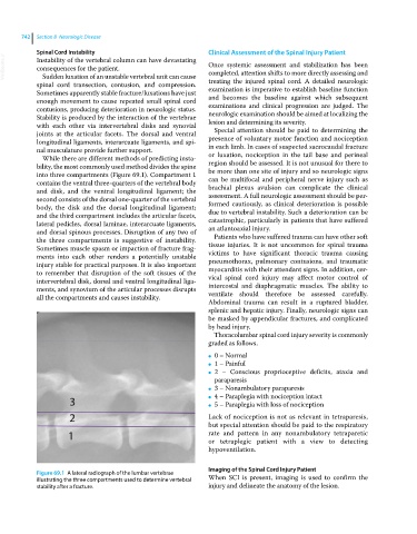
While there are different methods of predicting insta- or luxation, nociception in the tail base and perineal
bility, the most commonly used method divides the spine region should be assessed. It is not unusual for there to
into three compartments (Figure 69.1). Compartment 1 be more than one site of injury and so neurologic signs
contains the ventral three‐quarters of the vertebral body can be multifocal and peripheral nerve injury such as
and disk, and the ventral longitudinal ligament; the brachial plexus avulsion can complicate the clinical
second consists of the dorsal one‐quarter of the vertebral assessment. A full neurologic assessment should be per-
body, the disk and the dorsal longitudinal ligament; formed cautiously, as clinical deterioration is possible
and the third compartment includes the articular facets, due to vertebral instability. Such a deterioration can be
lateral pedicles, dorsal laminae, interarcuate ligaments, catastrophic, particularly in patients that have suffered
and dorsal spinous processes. Disruption of any two of an atlantoaxial injury.
the three compartments is suggestive of instability. Patients who have suffered trauma can have other soft
Sometimes muscle spasm or impaction of fracture frag- tissue injuries. It is not uncommon for spinal trauma
ments into each other renders a potentially unstable victims to have significant thoracic trauma causing
injury stable for practical purposes. It is also important pneumothorax, pulmonary contusions, and traumatic
to remember that disruption of the soft tissues of the myocarditis with their attendant signs. In addition, cer-
intervertebral disk, dorsal and ventral longitudinal liga- vical spinal cord injury may affect motor control of
ments, and synovium of the articular processes disrupts intercostal and diaphragmatic muscles. The ability to
all the compartments and causes instability. ventilate should therefore be assessed carefully.
Abdominal trauma can result in a ruptured bladder,
splenic and hepatic injury. Finally, neurologic signs can
be masked by appendicular fractures, and complicated
by head injury.
Thoracolumbar spinal cord injury severity is commonly
graded as follows.
0 – Normal
●
1 – Painful
●
2 – Conscious proprioceptive deficits, ataxia and
●
paraparesis
3 – Nonambulatory paraparesis
●
4 – Paraplegia with nociception intact
●
5 – Paraplegia with loss of nociception
●
Lack of nociception is not as relevant in tetraparesis,
but special attention should be paid to the respiratory
rate and pattern in any nonambulatory tetraparetic
or tetraplegic patient with a view to detecting
hypoventilation.
Imaging of the Spinal Cord Injury Patient
Figure 69.1 A lateral radiograph of the lumbar vertebrae
illustrating the three compartments used to determine vertebral When SCI is present, imaging is used to confirm the
stability after a fracture. injury and delineate the anatomy of the lesion.