
Page 970 - Clinical Small Animal Internal Medicine
P. 970
908 Section 9 Infectious Disease
Disease World Forum website (www.cvbd.org/en/ Frequently, pet owners will not recall tick exposure, since
VetBooks.ir occurrence‐maps/world‐map/). infection may have occurred weeks to months prior to
illness. Canine monocytic ehrlichiosis is more frequently
Recently, two new species of Ehrlichia have been
described in dogs in the United States: E. muris from a
sick dog in Minnesota, and Panola Mountain Ehrlichia associated with history of epistaxis and signs of pallor,
petechiation, and uveitis (Table 93.2, Figure 93.5). Canine
(PME) from a sick dog in North Carolina. E. muris is granulocytic ehrlichiosis and anaplasmosis are more fre-
believed to be transmitted by I. scapularis in the United quently associated with history of reluctance to stand or
States, with wild rodents as reservoir, whereas PME is walk and signs of joint effusion and lameness (see
transmitted by A. americanum, with white‐tailed deer as Table 93.2).
the probable vertebrate reservoir in the United States. Anaplasma platys infection rarely causes clinical signs
in the United States, but moderate to severe manifesta-
tion has been described in Europe, the Middle East, and
Signalment South America. Fever, lymphadenopathy, and spleno-
megaly are common findings in monocytic or granulo-
Male or female dogs at any age can develop the disease. cytic ehrlichiosis. Severe cases of monocytic ehrlichiosis
German shepherd dogs are predisposed to a more severe caused by E. canis may present with central nervous sys-
illness during E. canis infection. Despite the fact that cats tem (CNS) signs, retinal hemorrhage and detachment,
are naturally exposed to Ehrlichia and Anaplasma spp. and/or cardiac arrhythmias. The hallmark of the chronic
infection, clinical manifestations are rarely reported. phase of E. canis infection is pancytopenia from hypo-
plasia of all bone marrow cell lines, but protein‐losing
nephropathy, diffuse muscle wasting, and secondary
History and Clinical Signs infections, presumably due to immunosuppression, have
been described. Chronic illness associated with granulo-
The classic presentation in dogs is characterized by cytic ehrlichiosis/anaplasmosis has not yet been well
depression, lethargy, mild weight loss, and anorexia. documented in naturally infected dogs.
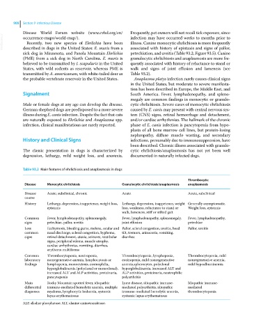
Table 93.2 Main features of ehrlichiosis and anaplasmosis in dogs
Thrombocytic
Disease Monocytic ehrlichiosis Granulocytic ehrlichiosis/anaplasmosis anaplasmosis
Disease Acute, subclinical, chronic Acute Acute, subclinical
course
History Lethargy, depression, inappetence, weight loss, Lethargy, depression, inappetence, weight Generally asymptomatic.
epistaxis loss, weakness, reluctance to stand or Weight loss, epistaxis
walk, lameness, stiff or stilted gait
Common Fever, lymphadenopathy, splenomegaly, Fever, lymphadenopathy, splenomegaly, Fever, lymphadenopathy,
signs petechiae, pallor, uveitis joint effusion petechiae
Less Ecchymosis, bleeding gums, melena, ocular and Pallor, scleral congestion, uveitis, head Pallor, uveitis
common nasal discharge, scleral congestion, hyphema, tilt, tremors, anisocoria, vomiting,
signs retinal detachment, ataxia, seizures, vestibular diarrhea
signs, peripheral edema, muscle atrophy,
cardiac arrhythmias, vomiting, diarrhea,
erythema multiforme
Common Thrombocytopenia, neutropenia, Thrombocytopenia, lymphopenia, Thrombocytopenia, mild
laboratory nonregenerative anemia, lymphocytosis or eosinopenia, mild nonregenerative nonregenerative anemia,
findings lymphopenia, monocytosis, eosinophilia, anemia,spherocytes, polyclonal mild hypoalbuminemia
hyperglobulinemia (polyclonal or monoclonal), hyperglobulinemia, increased ALT and
increased ALT and ALP activities, proteinuria, ALP activities, proteinuria, neutrophilic
pancytopenia polyarthritis
Main Rocky Mountain spotted fever, idiopathic Lyme disease, idiopathic immune‐ Idiopathic immune‐
differential immune‐mediated hemolytic anemia, multiple mediated polyarthritis, idiopathic mediated
diagnoses myeloma, lymphocytic leukemia, systemic immune‐mediated hemolytic anemia, thrombocytopenia
lupus erythematosus systemic lupus erythematosus
ALP, alkaline phosphatase; ALT, alanine aminotransferase.