
Page 971 - Clinical Small Animal Internal Medicine
P. 971
93 Ehrlichiosis and Anaplasmosis 909
Diagnosis
VetBooks.ir No single laboratory test is capable of detecting Ehrlichia
or Anaplasma infection in all cases, so clinicians should
be aware of the limitations of each diagnostic test
(Table 93.3).
Diagnosis is generally based on clinical and hemato-
logic abnormalities, response to therapy and presence of
specific antibodies against Ehrlichia and Anaplasma
spp. Common hematologic abnormalities are listed in
Table 93.2. In addition, it is worth noting that E. canis is
an important differential for lymphocytic leukemia or
multiple myeloma, as infection has occasionally been
associated with marked granular lymphocytosis and
monoclonal, rather than polyclonal gammopathy. E.
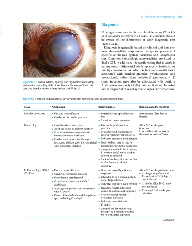
Figure 93.5 Corneal edema, opacity, and pigmentation in a dog canis infection may also be associated with positive
with uveitis caused by ehrlichiosis. Source: Courtesy of José Luis Antinuclear Antibody (ANA) tests, so it should be ruled
Laus and Ivan Ricardo Martinez Padua, UNESP, Brazil. out in suspected cases of systemic lupus erythematosus.
Table 93.3 Features of diagnostic assays available for ehrlichiosis and anaplasmosis in dogs
Assay Advantages Disadvantages Recommended testing time
Morulae detection ● Fast and cost‐effective ● Sensitivity and specificity are Acute phase (first days of
● Can be performed in practice low illness)
● Requires trained operator
IFA serology ● Gold standard, widely used ● Cannot be performed in After 2–3 weeks post
● Antibodies can be quantified (titer) practice infection.
Low antibody titers may be
● E. canis antigens cross‐react with ● Procedure not standardized detected as early as 7 days
Panola Mountain Ehrlichia among veterinary laboratories
● Can be used to monitor therapy ● Indicates exposure, not infection
(decrease in titers generally associated ● Four‐fold increase in titer is
with successful therapy) required for definitive diagnosis
● Assay not available for A. platys,
E. ewingii and E. muris as they
cannot be cultured
● Lack of antibody titer in the first
weeks does not rule out
exposure
ELISA serology: SNAP ● Fast and cost‐effective ● Does not quantify antibody After 2–3 weeks post infection
4Dx Plus ● Can be performed in practice response ● A. phagocytophilum and
● Procedure is standardized ● Intended to be a screening test, E. canis: after 7–21 days
post infection
not a diagnostic test
● E. canis spot cross‐react with E. A. platys: after 10–21 days
chaffeensis ● Indicates exposure, not infection ● post infection
● A. phagocytophilum spot cross‐react ● Negative results in the first E. ewingii: 3–4 weeks post
with A. platys weeks do not rule out exposure ● infection
● Detects five Ehrlichia and Anaplasma ● May not detect Panola
spp., including E. ewingii Mountain Ehrlichia
● Unknown sensitivity for
E. muris
● Limited use for monitoring
therapy, as it remains positive
for months after exposure
(Continued)