
Page 16 - Feline diagnostic imaging
P. 16
8 1 Digital Radiography
monitors range from 2 to 6 MP [7]. A 21 inch 3 MP monitor (a)
will commonly have a 2048 × 1526 pixel matrix. This level
of resolution approximates the average resolution of most
digital radiographs. This means that all the digital informa-
tion is displayed fully without having to magnify the image.
Although there is a clear advantage to medical‐grade
gray‐scale monitors, due to the expense it is difficult to jus- DENSITY
tify having them throughout the hospital. Consumer‐grade
color LCD monitors are generally considered adequate for
viewing images in exam rooms and surgery suites [7]. It is
advisable to have at least one medical‐grade gray‐scale
monitor in a dedicated reading area of the hospital for diag- LOG OF EXPOSURE
nostic interpretation of radiographs.
(b)
1.5 Comparison of Digital Versus
Analog Imaging
There are many advantages to digital imaging when com-
pared to traditional screen film systems such as reduced
expendable supply cost, consolidated image storage, and DENSITY
increased image portability and rapid referrals. In addi-
tion to these more obvious benefits, perhaps more impor-
tantly are the advantages in the image themselves. These
include exposure latitude and contrast along with image
processing [1].
With conventional radiography, the relationship LOG OF EXPOSURE
between exposure and optical density (blackness of the
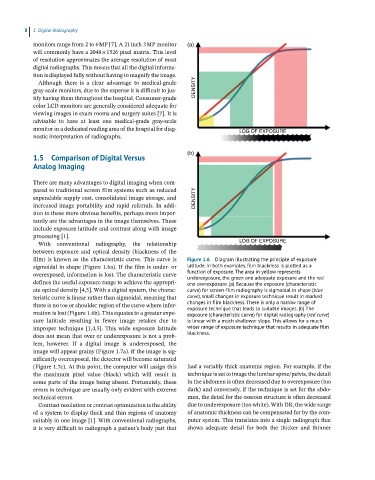
film) is known as the characteristic curve. This curve is Figure 1.6 Diagram illustrating the principle of exposure
sigmoidal in shape (Figure 1.6a). If the film is under‐ or latitude. In both examples, film blackness is plotted as a
overexposed, information is lost. The characteristic curve function of exposure. The area in yellow represents
underexposure, the green one adequate exposure and the red
defines the useful exposure range to achieve the appropri- one overexposure. (a) Because the exposure (characteristic
ate optical density [4,5]. With a digital system, the charac- curve) for screen-film radiography is sigmoidal in shape (blue
teristic curve is linear rather than sigmoidal, meaning that curve), small changes in exposure technique result in marked
there is no toe or shoulder region of the curve where infor- changes in film blackness. There is only a narrow range of
exposure technique that leads to suitable images. (b) The
mation is lost (Figure 1.6b). This equates to a greater expo- exposure (characteristic curve) for digital radiography (red curve)
sure latitude resulting in fewer image retakes due to is linear with a much shallower slope. This allows for a much
improper technique [1,4,5]. This wide exposure latitude wider range of exposure technique that results in adequate film
does not mean that over or underexposure is not a prob- blackness.
lem, however. If a digital image is underexposed, the
image will appear grainy (Figure 1.7a). If the image is sig-
nificantly overexposed, the detector will become saturated
(Figure 1.7c). At this point, the computer will assign this had a variably thick anatomic region. For example, if the
the maximum pixel value (black) which will result in technique is set to image the lumbar spine/pelvis, the detail
some parts of the image being absent. Fortunately, these in the abdomen is often decreased due to overexposure (too
errors in technique are usually only evident with extreme dark) and conversely, if the technique is set for the abdo-
technical errors. men, the detail for the osseous structure is often decreased
Contrast resolution or contrast optimization is the ability due to underexposure (too white). With DR, the wide range
of a system to display thick and thin regions of anatomy of anatomic thickness can be compensated for by the com-
suitably in one image [1]. With conventional radiographs, puter system. This translates into a single radiograph that
it is very difficult to radiograph a patient’s body part that shows adequate detail for both the thicker and thinner