
Page 307 - Feline diagnostic imaging
P. 307
312 18 Pleura
(b)
(a)
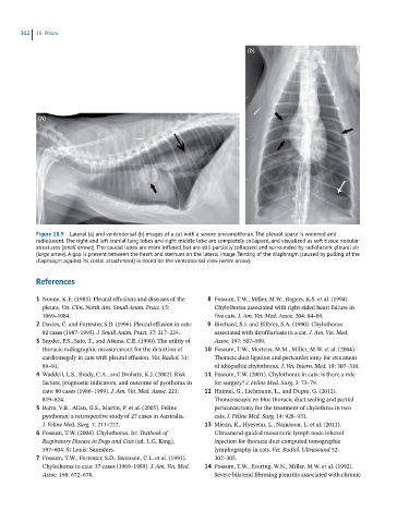
Figure 18.9 Lateral (a) and ventrodorsal (b) images of a cat with a severe pneumothorax. The pleural space is widened and
radiolucent. The right and left cranial lung lobes and right middle lobe are completely collapsed, and visualized as soft tissue nodular
structures (small arrows). The caudal lobes are more inflated, but are still partially collapsed and surrounded by radiolucent pleural air
(large arrow). A gap is present between the heart and sternum on the lateral image. Tenting of the diaphragm (caused by pulling of the
diaphragm against its costal attachment) is noted on the ventrodorsal view (white arrow).
References
1 Noone, K.E. (1985). Pleural effusions and diseases of the 8 Fossum, T.W., Miller, M.W., Rogers, K.S. et al. (1994).
pleura. Vet. Clin. North Am. Small Anim. Pract. 15: Chylothorax associated with right‐sided heart failure in
1069–1084. five cats. J. Am. Vet. Med. Assoc. 204: 84–89.
2 Davies, C. and Forrester, S.D. (1996). Pleural effusion in cats: 9 Birchard, S.J. and Bilbrey, S.A. (1990). Chylothorax
82 cases (1987–1995). J. Small Anim. Pract. 37: 217–224. associated with dirofilariasis in a cat. J. Am. Vet. Med.
3 Snyder, P.S., Sato, T., and Atkins, C.E. (1990). The utility of Assoc. 197: 507–509.
thoracic radiographic measurement for the detection of 10 Fossum, T.W., Mertens, M.M., Miller, M.W. et al. (2004).
cardiomegaly in cats with pleural effusion. Vet. Radiol. 31: Thoracic duct ligation and pericardectomy for treatment
89–91. of idiopathic chylothorax. J. Vet. Intern. Med. 18: 307–310.
4 Waddell, L.S., Brady, C.A., and Drobatz, K.J. (2002). Risk 11 Fossum, T.W. (2001). Chylothorax in cats: is there a role
factors, prognostic indicators, and outcome of pyothorax in for surgery? J. Feline Med. Surg. 3: 73–79.
cats: 80 cases (1986–1999). J. Am. Vet. Med. Assoc. 221: 12 Haimel, G., Liehmann, L., and Dupre, G. (2012).
819–824. Thoracoscopic en bloc thoracic duct sealing and partial
5 Barrs, V.R., Allan, G.S., Martin, P. et al. (2005). Feline pericardectomy for the treatment of chylothrax in two
pyothorax: a retrospective study of 27 cases in Australia. cats. J. Feline Med. Surg. 14: 928–931.
J. Feline Med. Surg. 7: 211–222. 13 Mieun, K., Hyeyeon, L., Namsoon, L. et al. (2011).
6 Fossum, T.W. (2004). Chylothorax. In: Textbook of Ultrasound‐guided mesenteric lymph node iohexol
Respiratory Disease in Dogs and Cats (ed. L.G. King), injection for thoracic duct computed tomographic
597–604. St Louis: Saunders. lymphography in cats. Vet. Radiol. Ultrasound 52:
7 Fossum, T.W., Forrester, S.D., Swenson, C.L. et al. (1991). 302–305.
Chylothorax in cats: 37 cases (1969–1989). J. Am. Vet. Med. 14 Fossum, T.W., Evering, W.N., Miller, M.W. et al. (1992).
Assoc. 198: 672–678. Severe bilateral fibrosing pleuritis associated with chronic