
Page 302 - Feline diagnostic imaging
P. 302
18.1 Pleural ffusion 307
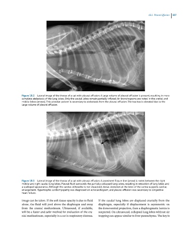
Figure 18.2 Lateral image of the thorax of a cat with pleural effusion. A large volume of pleural effusion is present, resulting in more
complete atelectasis of the lung lobes. Only the caudal lobes remain partially inflated. Air bronchograms are noted in the cranial and
middle lobes (arrows). This alveolar pattern is secondary to atelectasis from the pleural effusion. The trachea is elevated due to the
large volume of pleural effusion.
Figure 18.3 Lateral image of the thorax of a cat with pleural effusion. A prominent fissure line (arrow) is noted between the right
middle and right caudal lung lobes. Pleural fluid surrounds the partially collapsed lung lobes, resulting in retraction of lung lobes and
a scalloped appearance. Although the cardiac silhouette is not visualized, dorsal deviation at the level of the carina supports cardiac
enlargement. Hypertrophic cardiomyopathy was diagnosed on echocardiogram, and pleural effusion was secondary to congestive
heart failure.
image can be taken. If the soft tissue opacity is due to fluid If the caudal lung lobes are displaced cranially from the
alone, the fluid will pool above the diaphragm and away diaphragm, especially if displacement is asymmetric on
from the cranial mediastinum. Ultrasound, if available, the dorsoventral projection, then a diaphragmatic hernia is
will be a faster and safer method for evaluation of the cra- suspected. On ultrasound, collapsed lung lobes without air
nial mediastinum, especially in a cat in respiratory distress. trapping can appear similar to liver parenchyma. The key is