
Page 297 - Feline diagnostic imaging
P. 297
17.3 neumomediastinum 301
(b)
(a)
(c) (d)
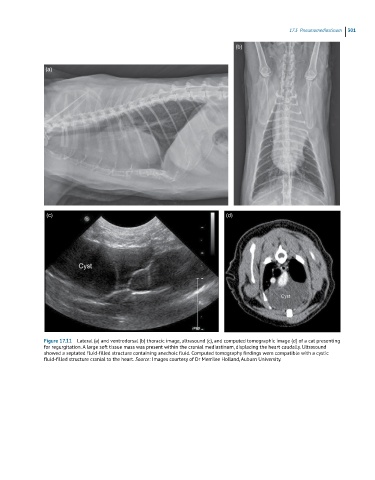
Figure 17.11 Lateral (a) and ventrodorsal (b) thoracic image, ultrasound (c), and computed tomographic image (d) of a cat presenting
for regurgitation. A large soft tissue mass was present within the cranial mediastinum, displacing the heart caudally. Ultrasound
showed a septated fluid-filled structure containing anechoic fluid. Computed tomography findings were compatible with a cystic
fluid-filled structure cranial to the heart. Source: Images courtesy of Dr Merrilee Holland, Auburn University.