
Page 292 - Feline diagnostic imaging
P. 292
296 17 Mediastinal Disease
(a) (b)
(c)
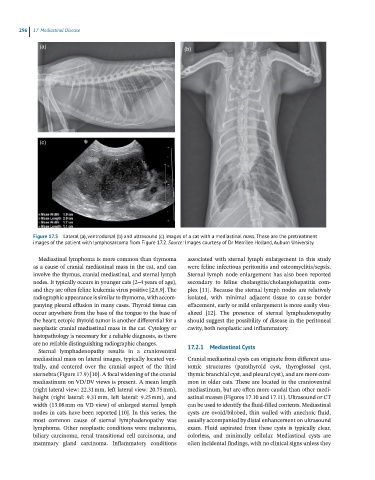
Figure 17.5 Lateral (a), ventrodorsal (b) and ultrasound (c) images of a cat with a mediastinal mass. These are the pretreatment
images of the patient with lymphosarcoma from Figure 17.2. Source: Images courtesy of Dr Merrilee Holland, Auburn University.
Mediastinal lymphoma is more common than thymoma associated with sternal lymph enlargement in this study
as a cause of cranial mediastinal mass in the cat, and can were feline infectious peritonitis and osteomyelitis/sepsis.
involve the thymus, cranial mediastinal, and sternal lymph Sternal lymph node enlargement has also been reported
nodes. It typically occurs in younger cats (2–4 years of age), secondary to feline cholangitis/cholangiohepatitis com-
and they are often feline leukemia virus positive [2,6,9]. The plex [11]. Because the sternal lymph nodes are relatively
radiographic appearance is similar to thymoma, with accom- isolated, with minimal adjacent tissue to cause border
panying pleural effusion in many cases. Thyroid tissue can effacement, early or mild enlargement is more easily visu-
occur anywhere from the base of the tongue to the base of alized [12]. The presence of sternal lymphadenopathy
the heart; ectopic thyroid tumor is another differential for a should suggest the possibility of disease in the peritoneal
neoplastic cranial mediastinal mass in the cat. Cytology or cavity, both neoplastic and inflammatory.
histopathology is necessary for a reliable diagnosis, as there
are no reliable distinguishing radiographic changes. 17.2.1 Mediastinal Cysts
Sternal lymphadenopathy results in a cranioventral
mediastinal mass on lateral images, typically located ven- Cranial mediastinal cysts can originate from different ana-
trally, and centered over the cranial aspect of the third tomic structures (parathyroid cyst, thyroglossal cyst,
sternebra (Figure 17.9) [10]. A focal widening of the cranial thymic branchial cyst, and pleural cyst), and are more com-
mediastinum on VD/DV views is present. A mean length mon in older cats. These are located in the cranioventral
(right lateral view: 22.31 mm, left lateral view: 20.75 mm), mediastinum, but are often more caudal than other medi-
height (right lateral: 9.31 mm, left lateral: 9.25 mm), and astinal masses (Figures 17.10 and 17.11). Ultrasound or CT
width (13.08 mm on VD view) of enlarged sternal lymph can be used to identify the fluid‐filled contents. Mediastinal
nodes in cats have been reported [10]. In this series, the cysts are ovoid/bilobed, thin walled with anechoic fluid,
most common cause of sternal lymphadenopathy was usually accompanied by distal enhancement on ultrasound
lymphoma. Other neoplastic conditions were melanoma, exam. Fluid aspirated from these cysts is typically clear,
biliary carcinoma, renal transitional cell carcinoma, and colorless, and minimally cellular. Mediastinal cysts are
mammary gland carcinoma. Inflammatory conditions often incidental findings, with no clinical signs unless they