
Page 296 - Feline diagnostic imaging
P. 296
300 17 Mediastinal Disease
(a) (b)
(c)
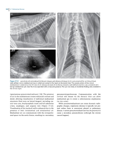
Figure 17.10 Lateral (a) and ventrodorsal (b) thoracic images, and ultrasound image (c) of a cat presented for vomiting. A focal,
well-defined cranial mediastinal mass is noted just cranial to the heart on the lateral view. The caudal portion of the cranial
mediastinum is widened on the VD view (*). An ultrasound exam of the thorax showed a well-defined anechoic cyst, consistent with
benign mediastinal cyst. Clear fluid was aspirated with ultrasound guidance. The cyst was likely an incidental finding, and unrelated to
the clinical signs.
(spontaneous pneumomediastinum) [20]. The presence pneumoretroperitoneum. Communication with the
of air in the mediastinum creates enhanced contrast and cervical soft tissues via the thoracic inlet can allow
detail, allowing visualization of individual mediastinal mediastinal gas to create a subcutaneous emphysema
structures (best seen on lateral images), including cra- (or vice versa).
nial vena cava, brachycephalic trunk and left subclavian While pneumomediastinum can create dramatic radio-
artery, esophagus, and azygous vein (Figure 17.12). graphic changes, respiratory distress is typically not pre-
Visualization of the tracheal wall is enhanced due to the sent unless there is concurrent pleural or pulmonary
presence of both intraluminal and extraluminal air. disease. A severe pneumomediastinum has the potential to
Mediastinal air can communicate with the retroperito - cause a secondary pneumothorax (although the reverse
neal space via the aortic hiatus, resulting in a secondary cannot happen).