
Page 291 - Feline diagnostic imaging
P. 291
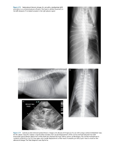
Figure 17.3  Ventrodorsal thoracic image of a cat with a mediastinal shift
               secondary to a unilateral pleural effusion. The heart is shifted (“pushed”) to
               the left because of increased volume in the right pleural space.


                     (a)                                                    (b)


                         (c)


               Figure 17.4  Lateral (a) and ventrodorsal (b) thoracic images and ultrasound image (c) of a cat with a large cranial mediastinal mass.
               On the lateral view, the trachea is significantly elevated, with caudal displacement of the carina and heart (beyond the sixth
               intercostal space). Border effacement of the heart and mediastinal mass mimics severe cardiomegaly. The cranial mediastinum is
               markedly widened on the VD view, again with caudal displacement of the heart. A complex, partially cystic mass is noted on the
               ultrasound image. The final diagnosis was thymoma.