
Page 286 - Feline diagnostic imaging
P. 286
290 16 Trachea
(b)
(a)
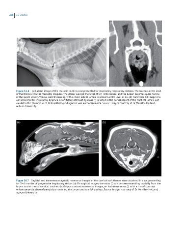
Figure 16.6 (a) Lateral image of the thoracic inlet in a cat presented for inspiratory respiratory distress. The trachea at the level
of the thoracic inlet is markedly irregular. The dorsal wall (at the level of C7) is thickened, and the lumen becomes quite narrow
at this point (arrow). Ventral wall thickening, with a more patent lumen, is present at the level of C6. (b) Transverse CT image of a
cat presented for inspiratory dyspnea. A soft tissue attenuating mass (*) is noted in the dorsal aspect of the tracheal lumen, just
caudal to the thoracic inlet. Histopathologic diagnosis was adenocarcinoma. Source: Images courtesy of Dr Merrilee Holland,
Auburn University.
(a) (b)
Figure 16.7 Sagittal and transverse magnetic resonance images of the cervical soft tissues were obtained in a cat presenting
for 5–6 months of progressive inspiratory stridor. (a) On sagittal images, the mass (*) can be seen extending caudally from the
larynx to the cranial cervical trachea. (b) On postcontrast transverse images, an isointense mass (*) with a rim of contrast
enhancement is circumferential surrounding the larynx and cranial trachea. Source: Images courtesy of Dr Merrilee Holland,
Auburn University.