
Page 285 - Feline diagnostic imaging
P. 285
16.4 Tracheal eoplasia 289
Figure 16.4 Lateral thoracic image of a cat presented with acute onset of coughing. A radiopaque foreign object is identified at the
carina. A small pebble was removed via bronchoscopy.
(b)
(a)
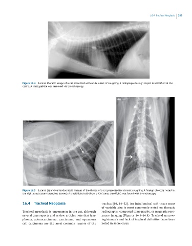
Figure 16.5 Lateral (a) and ventrodorsal (b) images of the thorax of a cat presented for chronic coughing. A foreign object is noted in
the right caudal stem bronchus (arrows). A small light bulb (from a Christmas tree light) was found with bronchoscopy.
16.4 Tracheal Neoplasia trachea [10, 16–22]. An intraluminal soft tissue mass
of variable size is most commonly noted on thoracic
Tracheal neoplasia is uncommon in the cat, although radiographs, computed tomography, or magnetic reso -
several case reports and review articles note that lym- nance imaging (Figures 16.6–16.8). Tracheal narrow-
phoma, adenocarcinoma, carcinoma, and squamous ing/stenosis and lack of tracheal definition have been
cell carcinoma are the most common tumors of the noted in some cases.