Page 280 - Feline diagnostic imaging
P. 280
284  15  Thoracic Cavity

                                (a)


                      (b)                                      (c)


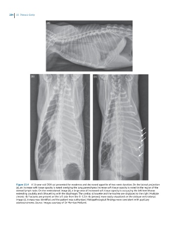
            Figure 15.4  A 16-year-old DSH cat presented for weakness and decreased appetite of two week duration. On the lateral projection
            (a), an increase soft tissue opacity is noted overlying the lung parenchyma. Increase soft tissue opacity is noted in the region of the
            sternal lymph node. On the ventrodorsal image (b), a large area of increased soft tissue opacity is occupying the left hemithorax
            extending caudally and silhouetting with the diaphragm. The cardiac silhouette and the trachea are displaced to the right. Multiple
            chronic rib fractures are present on the left side from the 8–12th rib (arrows) more easily visualized on the oblique ventrodorsal
            image (c). A mass was identified and the patient was euthanized. Histopathological findings were consistent with papillary
            adenocarcinoma. Source: Images courtesy of Dr Merrilee Holland.

