Page 278 - Feline diagnostic imaging
P. 278
282  15  Thoracic Cavity
                                                              15.1.2  Rib Neoplasia
                                                              Tumors  involving  the  ribs  are  one  of  the  most  common
                                                              sources of extrapleural mass lesions (masses that originate
                                                              outside the peripheral pleura). An extrapleural mass cre-
                                                              ates a broad‐based lesion with a convex margin facing the
                                                              lungs. The angle between the chest wall and mass should
                                                              be greater than 90°. These lesions typically grow inward
                                                              (toward the thoracic cavity) more than outward, and may
                                                              not be palpated or grossly visible. A secondary pleural effu-
                                                              sion  is  common  with  these  lesions,  and  the  chest  wall
                                                              should be evaluated with care in patients with pleural effu-
                                                              sion  of  unknown  etiology.  Computed  tomography  is  an
                                                              excellent imaging modality for a more detailed evaluation
                                                              of the ribs and pleural space in these cases.
                                                                The most common primary tumor affecting the ribs is
                                                              osteosarcoma. Other primary bone tumors occur rarely in
                                                              the  cat  (fibrosarcoma  and  chondrosarcoma),  with  no
                                                              reports of rib involvement [6,7]. Lysis and/or proliferation
                                                              are  common,  although  secondary  pleural  effusion  may
                                                              make visualization of the rib lesions more difficult early in
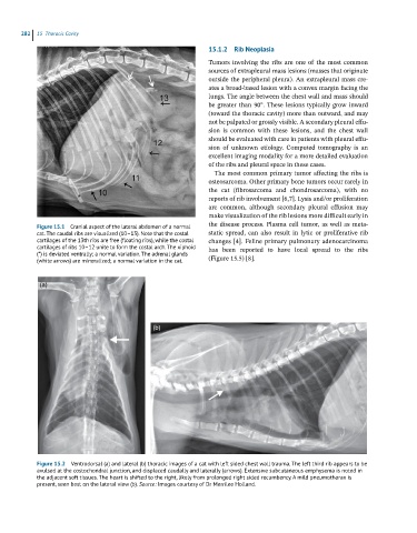
            Figure 15.1  Cranial aspect of the lateral abdomen of a normal   the disease process. Plasma cell tumor, as well as meta-
            cat. The caudal ribs are visualized (10–13). Note that the costal   static  spread,  can  also  result  in  lytic  or  proliferative  rib
            cartilages of the 13th ribs are free (floating ribs), while the costal   changes [4]. Feline primary pulmonary adenocarcinoma
            cartilages of ribs 10–12 unite to form the costal arch. The xiphoid   has  been  reported  to  have  local  spread  to  the  ribs
            (*) is deviated ventrally; a normal variation. The adrenal glands
            (white arrows) are mineralized; a normal variation in the cat.  (Figure 15.5) [8].


             (a)


                                              (b)


            Figure 15.2  Ventrodorsal (a) and lateral (b) thoracic images of a cat with left sided chest wall trauma. The left third rib appears to be
            avulsed at the costochondral junction, and displaced caudally and laterally (arrows). Extensive subcutaneous emphysema is noted in
            the adjacent soft tissues. The heart is shifted to the right, likely from prolonged right sided recumbency. A mild pneumothorax is
            present, seen best on the lateral view (b). Source: Images courtesy of Dr Merrilee Holland.

