Page 281 - Feline diagnostic imaging
P. 281
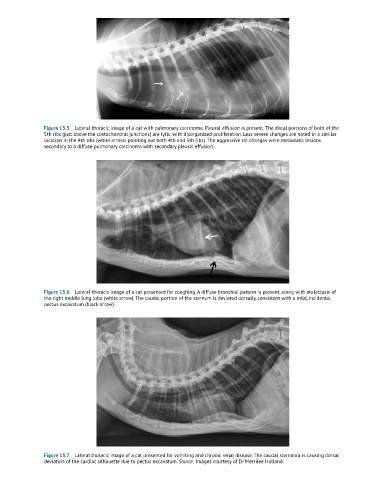
Figure 15.5  Lateral thoracic image of a cat with pulmonary carcinoma. Pleural effusion is present. The distal portions of both of the
               5th ribs (just above the costochondral junctions) are lytic, with disorganized proliferation. Less severe changes are noted in a similar
               location in the 4th ribs (white arrows pointing out both 4th and 5th ribs). The aggressive rib changes were metastatic lesions
               secondary to a diffuse pulmonary carcinoma with secondary pleural effusion.


               Figure 15.6  Lateral thoracic image of a cat presented for coughing. A diffuse bronchial pattern is present, along with atelectasis of
               the right middle lung lobe (white arrow). The caudal portion of the sternum is deviated dorsally, consistent with a mild, incidental
               pectus excavatum (black arrow).


               Figure 15.7  Lateral thoracic image of a cat presented for vomiting and chronic renal disease. The caudal sternabra is causing dorsal
               deviation of the cardiac silhouette due to pectus excavatum. Source: Images courtesy of Dr Merrilee Holland.

