Page 284 - Feline diagnostic imaging
P. 284
(b)


                                                            (a)


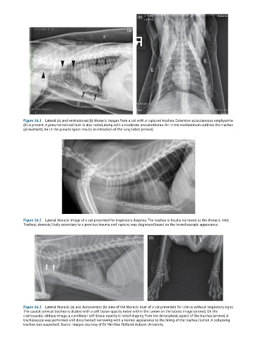
            Figure 16.1  Lateral (a) and ventrodorsal (b) thoracic images from a cat with a ruptured trachea. Extensive subcutaneous emphysema
            (E) is present. A pneumomediastinum is also noted, along with a moderate pneumothorax. Air in the mediastinum outlines the trachea
            (arrowheads). Air in the pleural space results in retraction of the lung lobes (arrows).


            Figure 16.2  Lateral thoracic image of a cat presented for inspiratory dyspnea. The trachea is focally narrowed at the thoracic inlet.
            Tracheal stenosis, likely secondary to a previous trauma and rupture, was diagnosed based on the bronchoscopic appearance.


                  (a)                                                 (b)


            Figure 16.3  Lateral thoracic (a) and dorsoventral (b) view of the thoracic inlet of a cat presented for icterus without respiratory signs.
            The caudal cervical trachea is dilated with a soft tissue opacity noted within the lumen on the lateral image (arrows). On the
            craniocaudal oblique image, a curvilinear soft tissue opacity is noted draping from the dorsolateral aspect of the trachea (arrows). A
            tracheoscopy was performed and documented narrowing with a normal appearance to the lining of the tracheal lumen. A collapsing
            trachea was suspected. Source: Images courtesy of Dr Merrilee Holland, Auburn University.

