Page 279 - Feline diagnostic imaging
P. 279
15.2 Diseisi of tsf sernu 283
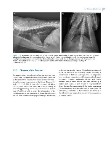
Figure 15.3 A nine-year-old DSH presented for inappetance. On the lateral image (a), there is a decrease in the size of the cardiac
silhouette. Focal air opacities are noted overlying the dorsal soft tissues and cranial abdomen. On the ventrodorsal image (b),
malalignment and fractures are noted in the ribs involving the sixth to tenth ribs on the right. Increase soft tissue and air are
noted in the right thoracic wall. Fluid opacity is present medial to the fractured ribs. Source: Images courtesy of
Dr Merrilee Holland.
15.2 Diseases of the Sternum pathology may also be present. Clinical signs are depend-
ent on the severity of the deformity, and are secondary to
Pectus excavatum is a deformity of the sternum and asso- compression of the heart and lungs. While some patients
ciated costal cartilages characterized by dorsal deviation have no clinical signs, others exhibit exercise intolerance,
of the sternebrae (usually the caudal sternebrae) and a tachypnea, cyanosis, respiratory distress, and cardiac
dorsal to ventral compression of the thorax (Figures 15.6 murmur. The murmur may be functional, secondary to
and 15.7) [9,10]. This is most often a congenital defect, cardiac malposition, or may be due to congenital heart
but acquired pectus has been described secondary to defect that may be concurrent with the pectus excavatum.
chronic upper airway resistance, with increased inspira- Clinical signs may be progressive, and in some cases, life
tory effort [9]. A mild to severe dorsal deviation of the threatening. Treatment is dependent on the severity of
caudal sternebrae with deviation of the cardiac silhouette the deformity, and ranges from conservative management
are the most common radiographic changes. Pulmonary to surgical repair.

