
Page 290 - Feline diagnostic imaging
P. 290
294 17 Mediastinal Disease
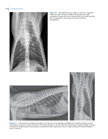
Figure 17.1 Ventrodorsal thoracic image of a cat with a mediastinal
shift to the right. The heart is shifted cranially and to the right
secondary to collapse and loss of volume of the right cranial lung lobe
(this is likely due to a mucus plug from chronic bronchial
inflammation).
(b)
(a)
Figure 17.2 Lateral (a) and ventrodorsal (b) images of the thorax on a cat undergoing treatment for mediastinal lymphoma. Fluid
accumulation is noted in the right hemithorax. The cardiac silhouette is shifted to the right which may be related to radiation therapy.
On the lateral image, fluid/soft tissue opacity is accumulated in the cranial thorax. Source: Images courtesy of Dr Merrilee Holland,
Auburn University.