
Page 298 - Feline diagnostic imaging
P. 298
302 17 Mediastinal Disease
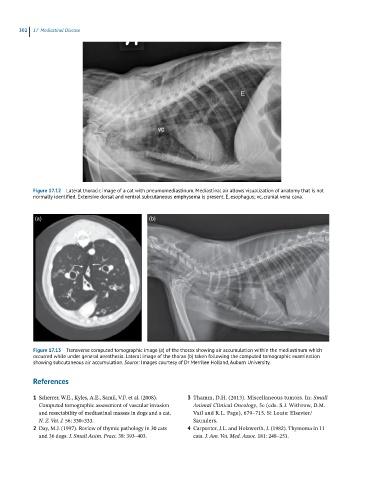
Figure 17.12 Lateral thoracic image of a cat with pneumomediastinum. Mediastinal air allows visualization of anatomy that is not
normally identified. Extensive dorsal and ventral subcutaneous emphysema is present. E, esophagus; vc, cranial vena cava.
(a) (b)
Figure 17.13 Transverse computed tomographic image (a) of the thorax showing air accumulation within the mediastinum which
occurred while under general anesthesia. Lateral image of the thorax (b) taken following the computed tomographic examination
showing subcutaneous air accumulation. Source: Images courtesy of Dr Merrilee Holland, Auburn University.
References
1 Scherrer, W.E., Kyles, A.E., Samii, V.F. et al. (2008). 3 Thamm, D.H. (2013). Miscellaneous tumors. In: Small
Computed tomographic assessment of vascular invasion Animal Clinical Oncology, 5e (eds. S.J. Withrow, D.M.
and resectability of mediastinal masses in dogs and a cat. Vail and R.L. Page), 679–715. St Louis: Elsevier/
N. Z. Vet. J. 56: 330–333. Saunders.
2 Day, M.J. (1997). Review of thymic pathology in 30 cats 4 Carpenter, J.L. and Holzworth, J. (1982). Thymoma in 11
and 36 dogs. J. Small Anim. Pract. 38: 393–403. cats. J. Am. Vet. Med. Assoc. 181: 248–251.