
Page 294 - Veterinary Immunology, 10th Edition
P. 294
VetBooks.ir
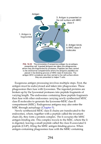
FIG. 10.10 The processing of exogenous antigen by an antigen-
presenting cell. Ingested antigens are taken into phagosomes
where they are fragmented by proteases. Peptides are then carried
to the endosomal compartments where the antigenic peptides are
placed in the binding grooves of MHC class II molecules. The
antigen-MHC complexes are then carried to the cell surface where
they are presented to helper T cells.
Exogenous antigen processing involves multiple steps. First, the
antigen must be endocytosed and taken into phagosomes. These
phagosomes then fuse with lysosomes. The ingested proteins are
broken up by the lysosomal proteases into peptide fragments of
varying length. The endosomes containing these peptide fragments
then fuse with other endosomes carrying newly synthesized MHC
class II molecules to generate the lysosome-MHC class II
compartment (MIIC). Endogenous antigens may also enter the
MIIC through autophagy (Chapter 5).
Newly synthesized MHC class II chains are translocated to the
endosomes, where, together with a peptide called the invariant
chain (Ii), they form a protein complex. The Ii occupies the MHC
antigen-binding site. This complex travels to the MIIC, where the Ii
is digested, leaving a small peptide called the class II-associated Ii
peptide (CLIP), filling the MHC antigen-binding groove. When
antigen-containing phagosomes fuse with the MHC-containing
294