
Page 356 - Veterinary Immunology, 10th Edition
P. 356
branches into penicillary arterioles. In some mammals, these
VetBooks.ir penicillary arterioles are surrounded by ellipsoids (periarteriolar
macrophage sheaths). These arterioles then open, either directly or
indirectly, into venous sinuses that drain into the splenic venules.
Ellipsoids are relatively large and prominent in pigs, mink, dogs,
and cats; are small and indistinct in horses and cattle; and are
absent in laboratory animals such as mice, rats, guinea pigs, and
rabbits. In species that lack ellipsoids, particles are trapped
primarily in the marginal zone of the white pulp.
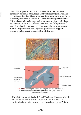
FIG. 12.19 Histological section and diagram showing the structure
of the bovine spleen. Original magnification ×50. (From a specimen
provided by Dr. J.R. Duncan.)
The white pulp contains both B and T cells, which accumulate in
their specific zones under the influence of chemokines. The
periarteriolar lymphoid sheaths consist largely of T cells. Within
356