
Page 627 - Veterinary Immunology, 10th Edition
P. 627
VetBooks.ir (the α chain of the IL-2 receptor). All activated T cells express CD25,
Treg cells are typical lymphocytes that express CD4 and CD25
but Treg cells are the only ones that express it when naïve. Their
most characteristic feature, however, is their use of a specialized
transcription factor called FoxP3. This is yet another example of a
situation in which cattle are different from mice and humans.
+
+
+
FoxP3 , CD4 , CD25 cells are found in cattle, but these are not
+
+
+
Tregs. The Treg cells in cattle are WC1.1 , WC1.2 , γ/δ T cells.
Natural Treg cells originate in the thymus (tTregs), whereas
peripheral Treg cells (pTregs) are produced in secondary lymphoid
organs, especially the intestine. The intestine is a major site of pTreg
development, and specialized intestinal dendritic cells promote this
through pathways that use a combination of transforming growth
factor-β (TGF-β), IL-2, and retinoic acid, a metabolite of vitamin A.
The retinoic acid is generated by the bacterial microbiota in the gut
and is required for normal T cell function. Intestinal pTreg cells
develop from naïve T cells in response to antigen and co-
stimulation. These signals induce the transcription of FoxP3. FoxP3
in turn induces transcription of the genes for CTLA-4, TGF-β, and
IL-10. pTregs are scattered throughout the body. They account for
roughly 5% of circulating T cells and 10% of lymph node T cells in
the dog.
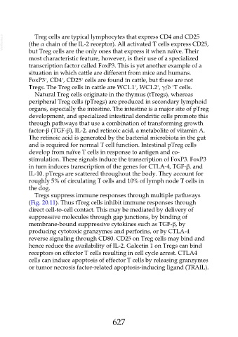
Tregs suppress immune responses through multiple pathways
(Fig. 20.11). Thus tTreg cells inhibit immune responses through
direct cell-to-cell contact. This may be mediated by delivery of
suppressive molecules through gap junctions, by binding of
membrane-bound suppressive cytokines such as TGF-β, by
producing cytotoxic granzymes and perforins, or by CTLA-4
reverse signaling through CD80. CD25 on Treg cells may bind and
hence reduce the availability of IL-2. Galectin 1 on Tregs can bind
receptors on effector T cells resulting in cell cycle arrest. CTLA4
cells can induce apoptosis of effector T cells by releasing granzymes
or tumor necrosis factor-related apoptosis-inducing ligand (TRAIL).
627