
Page 700 - Veterinary Immunology, 10th Edition
P. 700
species and among meals. The dog, for instance, has a low gastric
VetBooks.ir pH relative to that of the pig. Similarly, the pH in the center of a
mass of ingested food may not necessarily drop to low levels, and
some foods such as milk are potent buffers. In addition to
antimicrobial peptides, lysozyme is synthesized in the gastric
mucosa and in macrophages within the intestinal mucosa. As a
result, it is found in large quantities in intestinal fluid.
In the small intestine, separation of the microbiota and
enterocytes is maintained by a layer of mucus containing multiple
antimicrobial proteins. In the large intestine this separation is
maintained by two distinct layers of mucus. The inner layer is
almost bacteria free. The outer loose layer contains large numbers
of bacteria. In the developing animal it takes time for these mucus
layers to develop, and this provides a window of opportunity for
organisms such as segmented filamentous bacteria (SFB) to reach
and bind enterocytes.
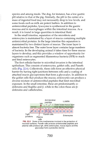
The first cellular barrier to microbial invasion is the intestinal
epithelium. This consists of enterocytes, goblet cells, and Paneth
cells (Fig. 22.6). Collectively, these cells form an effective physical
barrier by having tight junctions between cells and a coating of
attached mucin glycoproteins that form a glycocalyx. In addition to
the goblet cells that produce the mucus, enterocytes can produce a
diverse mixture of antimicrobial peptides that limit microbial
exposure. In the small intestine, these are predominantly α-
defensins and RegIIIα and β, while in the colon these are β-
defensins and cathelicidins.
FIG. 22.6 Some of the mechanisms involved in the protection of
mucosal surfaces. Paneth cells produce antimicrobial peptides and
plasma cells produce IgA, while enterocytes, mucus layers and the
glycocalyx form a protective barrier.
700